Book review: Manual of Tree Statics and Tree Inspection

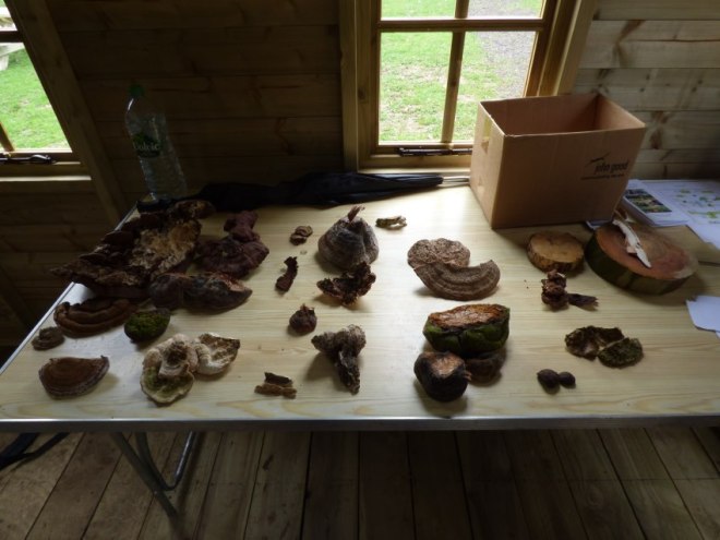
First and foremost, for those interested in purchasing the English version of the book, you can do so via the publisher Patzer Verlag, Summerfield Books or the Arboricultural Association. It’s not a cheap book and thus, for many, demands strong consideration before purchase, which is one of the core tenets behind why I wanted to read and review this book promptly following on from its publication. Therefore, my intention with this review is to give more information about the book over and above simply the listing of the chapters and sub-chapters, which are provided by the book sellers. Hence, interspersed amongst my review are photographs of the book, which will hopefully help guide you in your decision-making process. In fact, here’s an image for you, off the bat!

P1270705
Quite a nicely presented hardback. The select illustrations you see on the front here occur throughout the book – hardly a page goes by where there isn’t at least one such image.

As regards the content, therefore, the book begins with a very succinct introduction followed by an extensive assessment of the countless aspects pertaining to tree biology. This biology section is adorned with illustrations that, as is the case across the entire book, help greatly with the simple conveyance of the core messages put forth by the authors – indeed, as is so prevalent and well-received across Mattheck’s works (in spite of the disagreements the autors have with the t/r ratio and h/d ratio, which they detail within). Somehow, I doubt this inclusion of illustrations is coincidental and instead a homage or emulation of what works well when describing detailed concepts in only a few words. Critically, with a book detailed as one that covers tree statics, the incorporation of tree biology into the statics model is included, which keeps the book on track from the beginnings and saves it from veering off course into just one that echoes prior understanding.

At this point, an interjection is necessary. Specifically, for the purpose of highlighting some of the very curious qualities of this chapter – in the good sense. Principally, an addition to the developing understanding of summer branch drop is provided, whereby the authors detail that, on top of the impact drought has on the failure of large branches, temperature plays a possibly significant role. Why, you ask? Because, as the authors detail, the pretensioning of a tree’s wood fibres (i.e. the manner in which it loads itself under its own weight and optimises itself for this) is lessened by heat, which relaxes the wood fibres – including atop branch junctions. When compiled with the fact that trees are much stronger under tension than compression, the developing compression zones underneath the branch junction (and just behind the branch collar) as the wood fibres relax atop build up and, alongside other factors (including drought and probably many other unknown variables), the branch can subsequently suddenly fail. The authors delve into this phenomenon more in the third chapter and beyond. Other intriguing and enlightening aspects of this second chapter include the remarks on how construction works impact upon tree stability, in addition to the efficacy of ivy to be both a bane and a blessing for the tree, as regards oscillation and damping during wind loads.

It is however the third chapter that arguably provides the most appreciable benefit to the reader, as it takes us into the realm of tree statics in the direct sense. The intricacies of the chapter will, of course, largely evade this review, as the intention of this post is not to ‘spoil’ the contents of the book to any marked degree and make its purchase redundant. Instead, what can be said is that it delves effectively into critical facets of statics and, in my personal opinion, the segment on wind loading and the impact of crown architecture and elevation on loading forces is most brilliant – even down to the illustrations and mathematical examples, which really do simplify an evidently complex engineering approach to tree risk assessment.

P1270711
A segment from this third chapter, demonstrating how the authors blend text with diagrammatic models and more general illustrations.

Moreover, the discussion on the importance of hollowness for trees and why it should not always be considered a concern to the tree inspector is welcomed (unless it’s an open cavity and then, please, do be more worried straight off of the bat). In fact, and I quote: “the dimensions of a cavity in a tree have absolutely no informative value, as long as the occurring loads are not known.” Therefore, the t/r ratio Mattheck provided, the authors allege, is meaningless and incorrect.

Amazingly (from a factoid perspective), the authors then detail that a tree with a dbh of 1m and a central cavity leaving only 10cm of sound radial wood around the circumference has the same load-bearing capacity as a tree that is completely sound and has a dbh of 84cm! Of course, from an angle of understanding where the maximum loads occur, the fact that wood fibres stretch and compress far more at the outer sections of the stem than they do internally (with the neutral axis generally being the centre) gives credence and context to this assertion. The understanding of hollowness on older (veteran) trees is also discussed in a very articulate manner and, as it so seems, the greater risk for such trees is not failure of the trunk or root plate but of large lateral limbs attached to a hollowing trunk that can no longer sufficiently support such a mechanical load.

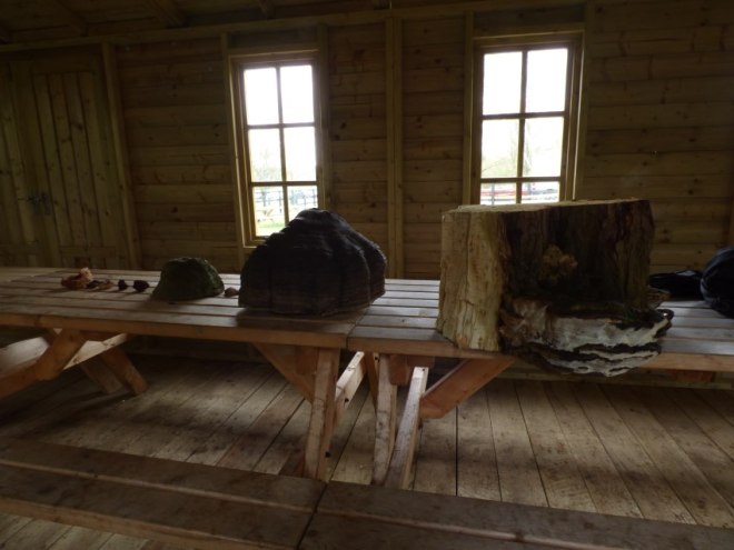
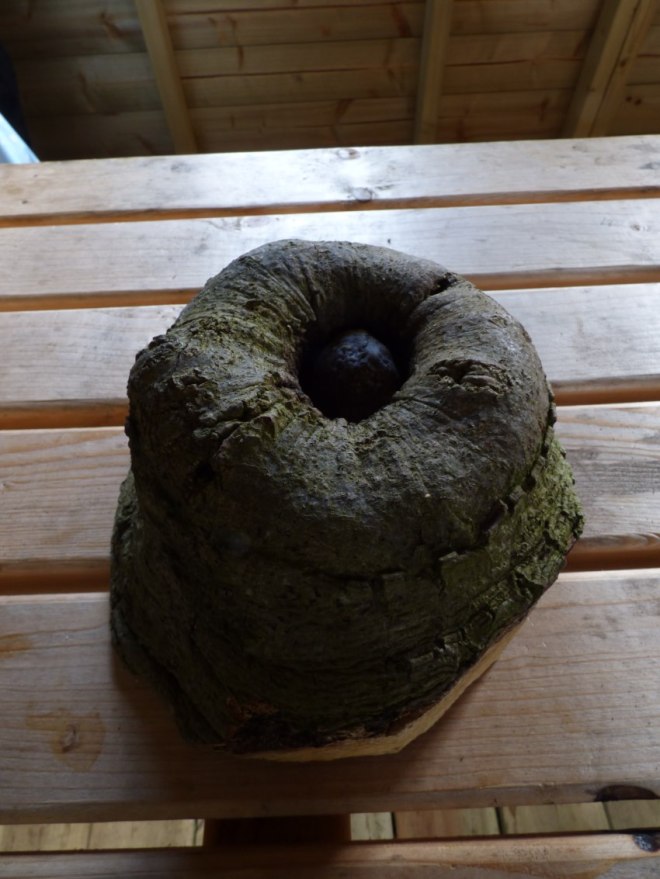
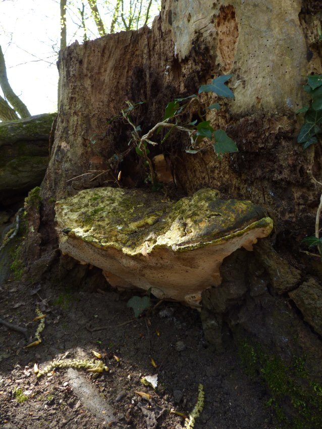
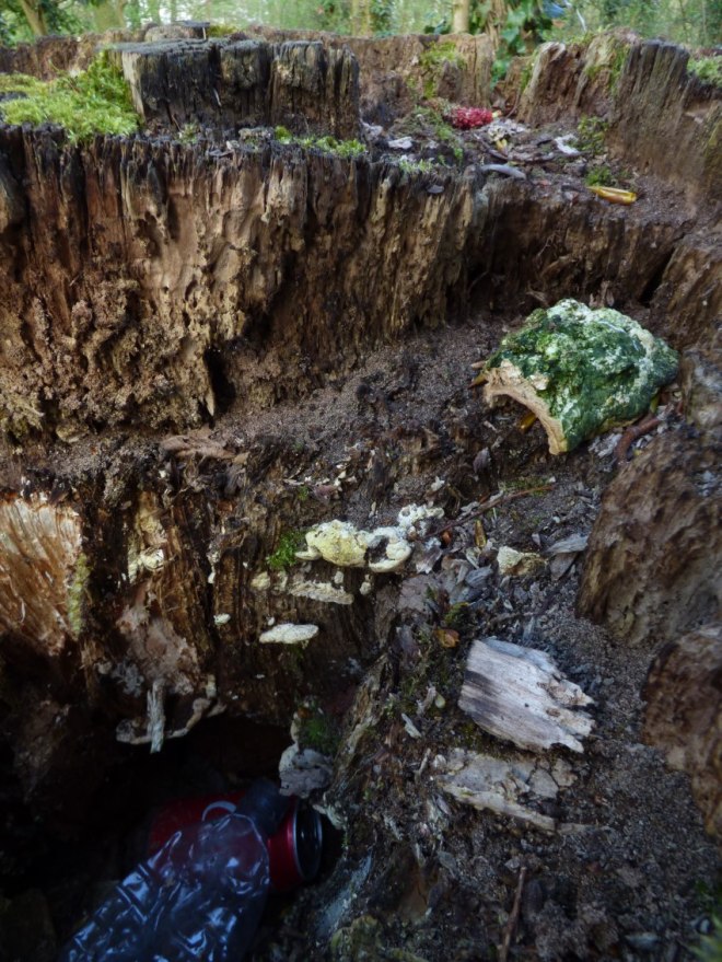
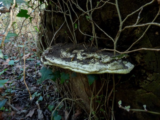
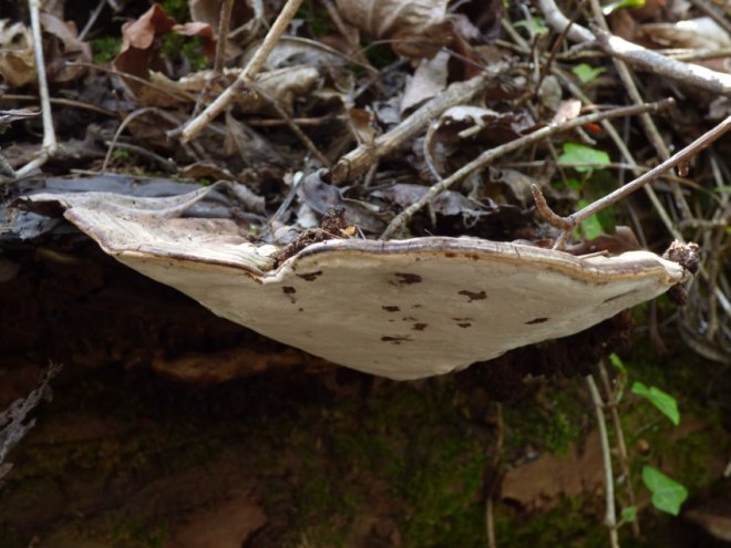
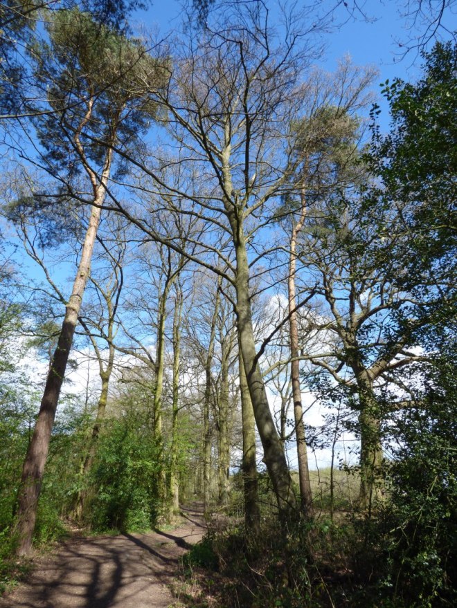
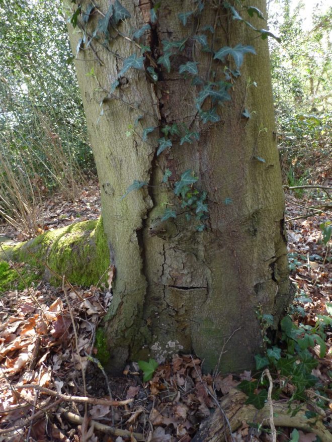
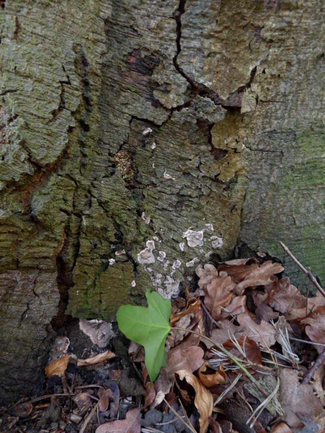
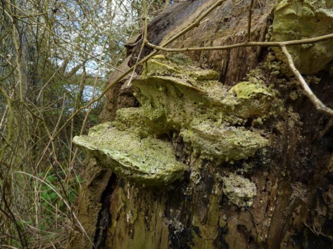
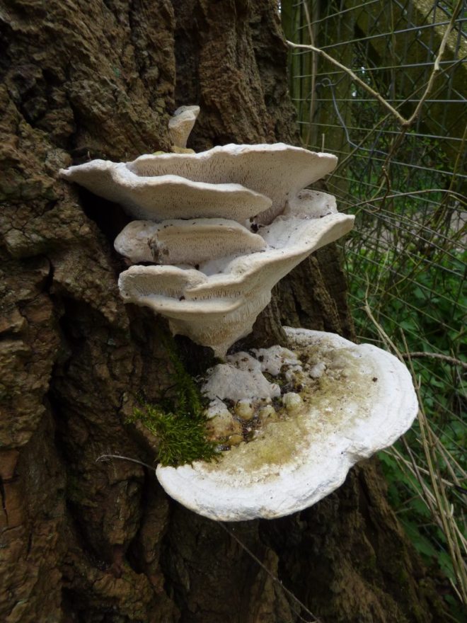
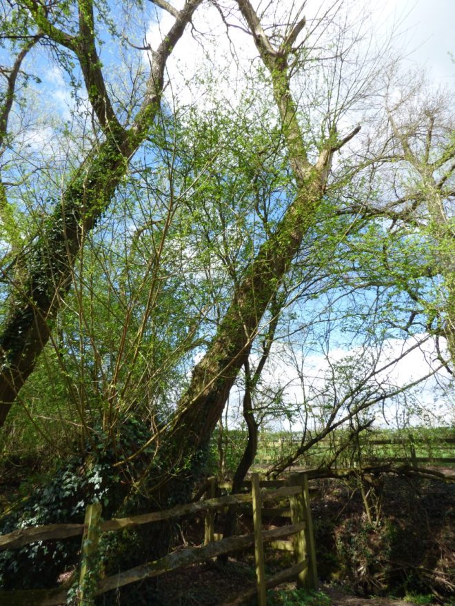
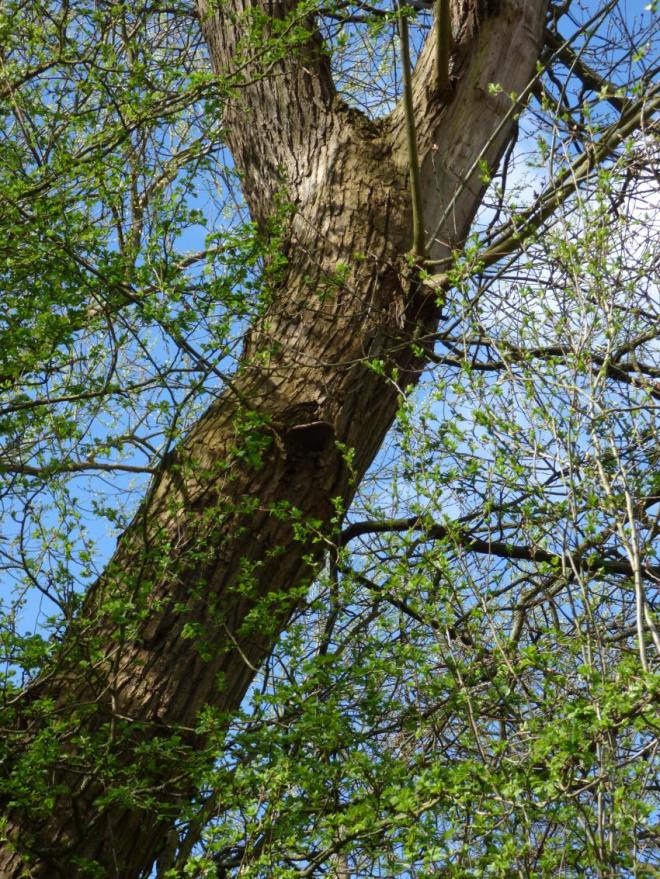
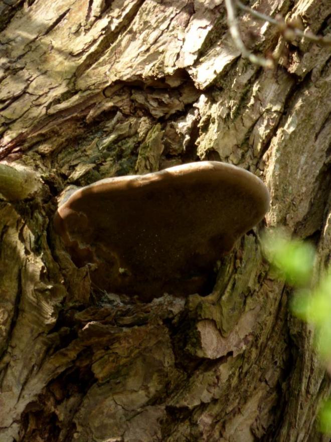
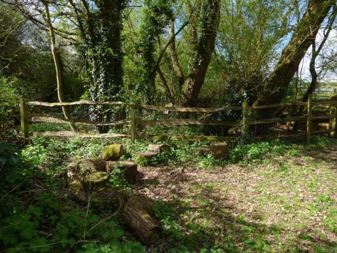
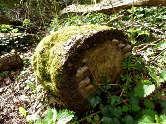
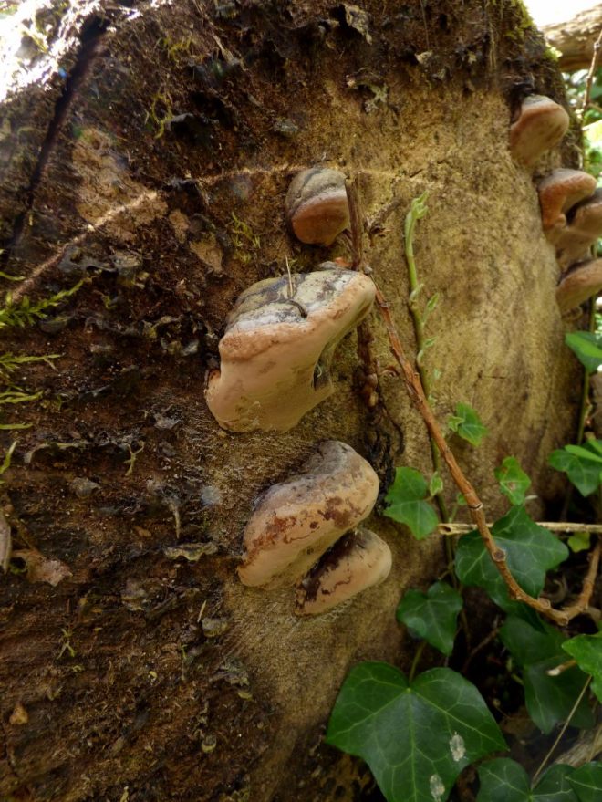
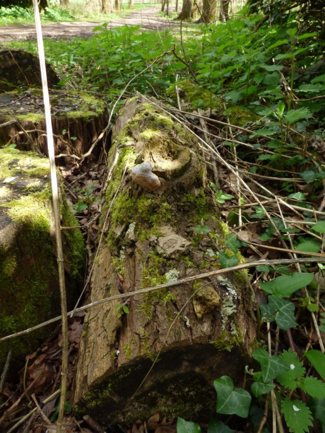
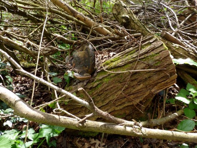
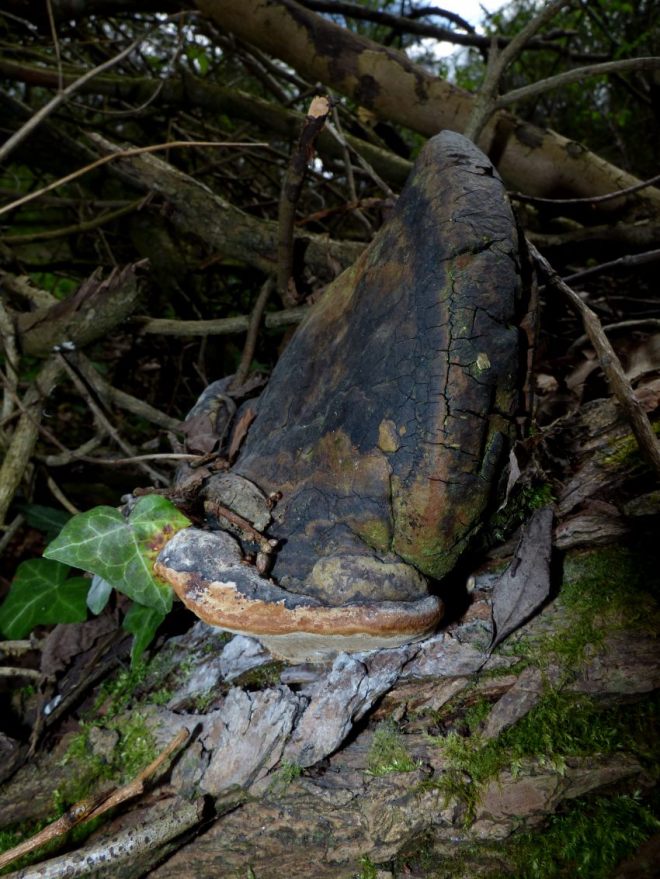
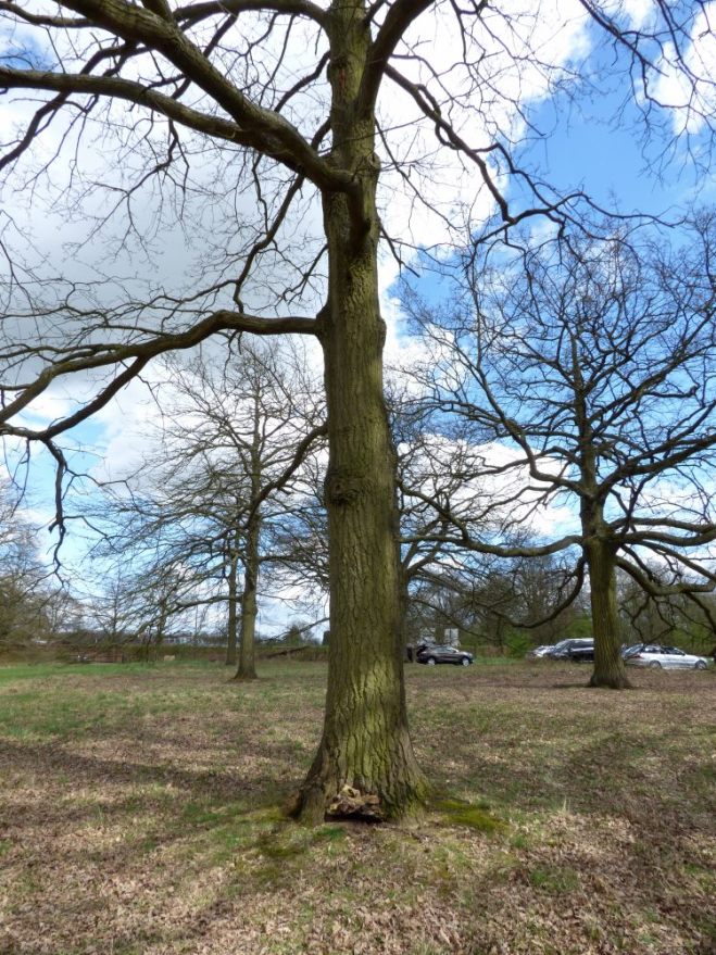
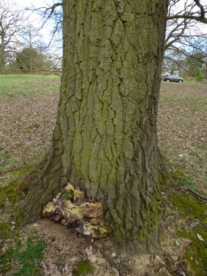
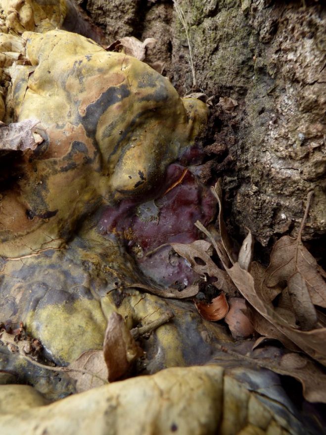
On this note, the outlining of root plate architecture is also deserving of mention. In this third chapter, the authors do a superb job of explaining why and how root plates fail and why, for all intents and purposes, extensive buttressing and adventitious rooting is not necessarily to be looked upon as defective. Using the example of a mature beech (see a below photo that I took recently), they allude to the often very pronounced root plate acting as a counter-balance to the lever arm that is the trunk (think of a very wide-based wine glass and compare it to a narrow-based one – which tilts more readily?). Indeed, where fungal decay is evident, as in the below photo, the effectiveness of the wide root plate comes into question, however.

Fagus sylvatica mature buttressing Ganoderma Meripilus 8
The admirable root plate of a mature beech. In this case, Ganoderma australe is compromising the butt and principal rooting structure, though where decay is not evident or is limited then a wider root plate is very effective at supporting the tree during times of wind loading – certainly more so than where such a wide root plate is not evident, on mature beech.

At this point, the notion of slenderness also enters the equation and, very curiously, the book asserts that slender multi-stemmed trees are more likely to oscillate excessively in wind loading conditions and fail, when compared to single-stemmed trees of equal slenderness. However, more crucially, why slenderness equals higher oscillatory frequency and thus denotes a greater risk of failure is detailed, which drives home the importance of not thinning out groups of trees and expecting the remaining ones to be able to stand wind-firm (the so-called ‘domino effect’ is also defined, whereby trees at a woodland edge successively fail, as those around become exposed after an initial tree fails and then those around it also fail).

And my favourite quote(s) of the third chapter? In reference to reaction growth laid down by the tree due to decay or other structural matters: “the development of symptoms is an expression of the vitality of the tree, not of its weakness” and that “focussing on so called symptoms can be absolutely counterproductive” – assuming, of course, you don’t quantify the weakness by ascertaining its load-bearing capacity (which is where statics comes in). Assumptions, therefore, are not good (though we all do assume, every day, when we assess trees, when working with outward symptoms solely).

P1270710
A segment of the fourth chapter. This double page spread is certainly one of the rarer examples of where illustrations do not dress the paper in abundance.

Moving into the fourth chapter, we’re first greeted with a rather novel take on tree assessment: do not negate the emotional aspect of the act. To be precise, from this, I understand it as the authors inferring that the intuitive assessment of a tree, from afar, takes into account both our logical deduction of its form (based on the scientific understanding of what denotes good structural architecture in trees) and surrounds (targets, exposure, etc) and also its innate emotional ‘appeal’ (i.e. is the tree ‘harmonious’ and flowing well, or is it a rough and jagged structure that is abrasive to the eye and clearly there is something awry that demands more investigation). Here, the authors then throw in the concept of a female breast being harmonious (and not at all like a brick…!) to a child, who can appreciate the allure of that particular aspect of female anatomy for its biological purposes. I presume this wasn’t a translation error (of which there are a few, across the entire book, but it largely flows very well)! Thus, trees are like female breasts: harmonious (I could end the review here, quite frankly, for the comedic value).

Anyway, this fourth chapter then goes on to outline, to a very detailed degree, with the support of various diagrams in the sixth chapter (the ‘Annexes’), more about SIA (static integrated assessment). For something that I admittedly had considered quite complex, which it still certainly is, the authors do a stellar job in simplifying the concept and utilise diagrams and drawings to help ensure the reader can understand the message being conveyed. The explanation of the rationale behind why this methodology, provided in preceding chapters and also in this one, helps to contextualise the methodology and, quite truthfully, it’s a form of tree assessment that I am now keen to put into some form of practice. The statement the authors make about microdrills essentially being redundant now that sonic tomography has arisen from the mulch is also a curious one, which is deserving of some consideration, when appreciating that the former causes damage to trees and the latter, almost wholly, does not; in addition to the ability for a tomograph (i.e. PiCUS or ARBOTOM) to plot an entire cross-section and not just track the path of a single drill bitand provide information from that sole path.

A further useful segment within the fourth chapter is that of the pests and pathogens of the leaves and shoots of many common European tree species and genera. Indeed, the assertion that horse chestnut leaf miner is not solely an aesthetic issue is a welcome one, as for whatever concerning reason that understanding of it being only an aesthetic pest is still accepted by some in the industry. The section also has a really nice little bit of information on Massaria disease of plane, with a distinct Teutonic angle (as one would expect, from German authors).

P1270725
A double page spread of the section of pests and diseases of the shoots and leaves of trees common to Europe.

The (‘technically’) last chapter, the fifth, draws upon the information provided within all prior chapters and provides the reader with guidance on how to, following the identification of a need to manage the tree (be it a newly-planted tree or veteran in a park or construction site), effectively enact a management regime that will be of benefit to the longevity of the tree in the landscape (this is why we manage many trees in ways that doesn’t see them get felled). Indeed, this chapter presents a lovely succinct look at the need to strongly consider management options, as in deciding upon a route there is then, in theory, no going back upon that decision (once scheduled work has been completed).

The encouragement by the authors to refrain from using a rigid support system, be that system a stake for a young tree or steel braces for old trees with slender co-dominant limbs, and instead utilise a flexible and dynamic system, is therefore well-received, as they present their argument in a sound and logical manner. Importantly, for bracing, the authors even go so far as to detail what angle a brace should be applied at and the load that it should be able to bear relative to the size of the parts being connected, which elevates this book to a level beyond that of others that, from memory, do not provide as much detail. Their comments on propping with A-frames and not upside-down V-frames are also welcomed and articulated so well with so few words. Again, illsurations really help instruct the reader and break the text apart, thereby making the reading more ‘bite-sized’.

limb propping tree lateral load a-frame
This duo of props fails to suppprt the limb against lateral forces, given their design. In lacking an A-framed design, these props are thus not optimally positioned to keep the limb of this sycamore from failing under a torsional load.

Finally, the appendices presented in chapter 6, which support many of the assertions made in the earlier chapters, arrive. There exist some fabulous tables showcasing the wood properties of different tree genera (cellular make up, strength, etc), the speed at which tree genera move water up the trunk in metres per hour, the compartmentalisation ability of different species and genera, and a comparison of various forms of tree assessment and their strengths / weaknesses. Also within these annexes is information on how hollow trees fail and a description of principal wood-decay fungi and their significance. The fungal section is limited in detail and species diversity (because it’s only an annex and thus cannot possibly be as detailed as a dedicated book on fungi) though, concerningly, the brittle cinder (Kretzschmaria deusta) is described as a white rot in a comparitive table (whilst being correctly marked as a soft rot in its descriptive text). Proofing of the text should have picked this up, as it is perhaps the most significant mistake in the book (all other issues identified are largely translation errors or typos, from what I could spot). The comments on Perenniporia fraxinea preferring robinia (Robinia pseudoacacia) over any other host tree in urban areas where soil is compacted are, nonetheless, very interesting. Anecdotally, I can understand and agree with this assertion.

So, my thoughts to summarise? This is a very useful book that serves not only as a primer for the statics integrated assessment model but also for tree visual assessments on the whole. The eagerness of the authors to promote analytical thinking of the reader was pervasive throughout and, in some respects, this book could essentially serve as a 101 for tree inspections and management to any tree inspector. Of course, the title alludes to this by referring to tree inspection, though I admit I was taken aback (in a positive sense) on how much breadth this book covered in so few words and so many images. For me, the mix works well and therefore the looming threat of a wall-of-text was evaded. Perhaps it is just me, though I can pick up influences from Shigo and Mattheck, in the use of few choice words to describe complex matters and to support such words with harmonious (see what I did there?) images.

If you have around £115 to spend and want a book that you can refer to routinely, this will be worth purchasing. Don’t think you’ll be getting an impossibly wordy book that delves into tree statics down to the minute level, however. That is reserved for other literature and, in my perspective, rightly so.

Book review: Manual of Tree Statics and Tree Inspection

A trip to Aldenham Country Park – trees and fungi

With the weather remaining fair, in spite of the onerous musings spouted from the verbal orifices of the meteorological office, getting out at the weekend to explore new sites is still very much on the cards. Today, a group of us went over to Aldenham Country Park in north-west London, to search for interesting fungi on trees; as if a weekend would yield any other result!

We started the day by doing something socially reprehensible: bringing in fungi collections for display. As the below photos show, my collection is growing in extent, though is dwarfed in literal size by another collection, which essentially involves monster brackets that are, in some cases, still clinging to the very substrate that provided them with their life.

polypore collection fungi
My collection, consisting of fruiting bodies of fungi including the genus Ganoderma (top left), the genus Trametes (bottom left), Fomes fomentarius (top middle), the genus Phellinus (bottom right) and Coriolopsis gallica (bottom right).
polypore collection 2
Another collection, set up almost like a demonstration of the solar system (with the Perenniporia fraxinea on the poplar being the sun, of course!), including Fomes fomentarius (a monster one), Daedalea quercina and, as stated, the Perenniporia fraxinea on the poplar wood.

Before sharing some finds from today, it’s almost important to share some images of more cross-sectional decay as caused by Ganoderma pfeifferi. For those of you with a memory that stretches back beyond a mere seven days, you might recall a recent post I made showing a decay cross-section on a failed beech. Below, we see how the fungus’ activity within a branch stub of a beech has resulted in zonal decay, which is somewhat comparable to the other example shared recently – particularly, with regards to the rosing pattern.

Ganoderma pfeifferi internal decay 1
A tiny Ganoderma pfeifferi within the opening of a branch stub wound on beech.
Ganoderma pfeifferi internal decay 2
The cross-section of decay produced by the fungus.

And so, on with the walk we did, quite early on we wandered past an old poplar stump with some quite extensive Rigidoporus ulmarius decay. Indeed, as is quite routine with this fungus, the internal hollow was clad aplenty with small brackets, whilst the outside sported a much more sizeable fruiting body still in an active phase of its existence. Evidently, a new hymenium has recently been laid down, suggesting that this fungus is soon ready to begin producing spore for the coming season.

Populus Rigidoporus ulmarius stump decay 1
Rigidoporus ulmarius acting as a saprotroph on this senescent stump.
Populus Rigidoporus ulmarius stump decay 2
Quite a nice one, actually! Good morphology.
Populus Rigidoporus ulmarius stump decay 3
Looking inside the hollow, not only can we see that it is used as a bin, but also to house many small fruiting bodies of this fungus.

Very soon after this sighting, a fallen poplar log with Oxyporus populinus was discovered. I admit to only having seen this fungus twice, of which this find was one, so for me this was particularly exciting. In fact, the single fruiting body was rather massive and easily discernible by the quite brilliant tube layers separated by narrow bands of mycelium. Almost directly adjacent to this was a fruiting body of Ganoderma applanatum, as could be determined morphologically by the very thin cuticle atop the bracket (that is crushed easily and cuts very easily) and the extensive damage to the fruiting body, as caused by the yellow flat-footed fly Agathomyia wankowiczii.

Oxyporus populinus Populus log 1
A poplar log hides amongst ivy.
Oxyporus populinus Populus log 2
On one of the cut ends sits this large fruiting body of the fungus Oxyporus populinus.
Oxyporus populinus Populus log 3
The demarcations between each growth spurt are incredibly distinct, in this fungus.
Ganoderma applanatum Populus log 1
A fruiting body of Ganoderma applanatum also sat nearby, on the same log.
Agathomyia wankowiczii Ganoderma applanatum Populus log 3
Underneath, we can see the distinct gall structures caused by the yellow flat-footed fly.
Agathomyia wankowiczii Ganoderma applanatum Populus log 2
We can also see the internal damage caused by the fly, as it develops into its adult form and leaves to lay eggs elsewhere. The very thin upper cuticle can also be seen, which is thicker on Ganoderma australe.

Following the sighting of copious amounts of Daedaleopsis confragosa, our attention was then drawn to a rather sorry-looking beech tree over a well-used footpath. Upon close inspection, both Kretzschmaria deusta and the rhizomorphs of Armillaria mellea could be found, which certainly puts the longevity of this beech as is into doubt. To be honest, in all likelihood it’ll be monolithed, in order to still provide habitat but with the risk removed.

Kretzschmaria deusta beech Fagus Armillaria 1
It even leans over the footpath!
Kretzschmaria deusta beech Fagus Armillaria 2
Both the anamorphic stage of Kretzschmaria deusta and cambial necrosis caused by Armillaria mellea can be seen, in this image.
Kretzschmaria deusta beech Fagus Armillaria 3
Not looking good for this beech!

Around the proverbial corner (it was more like a ten minute trundle) from this beech stood a massive stump of an old poplar. In its prime, this would have been a tree operating on beast-mode, though is now far more modest in size. However, to make up for its literal demise, it now is host to the fungus Trametes gibbosa, which can be seen around one of the two stems.

Trametes gibbosa Populus stump 1
A fortress of nettles guards this poplar stump.
Trametes gibbosa Populus stump 2
Too bad they can’t defend against a zoom lens and / or walking boots and jeans!
Trametes gibbosa Populus stump 3
Some fresh brackets adorn the opposite side of the stump.
Trametes gibbosa Populus stump 4
Quite pretty, to be honest!

Delightfully, this stump also housed a bird nest, which I found only by pure chance when noticing what looked like chocolate mini-eggs! Tucked away impossibly well within a bark crevice was a small robin’s nest (I think), complete with four eggs. Hopefully, this stump will offer enough privacy to enable the chicks to develop well and not get picked-off by predators.

Erithacus rubecula eggs poplar stump tree 1
The arrow shows where the nest is, as it’d otherwise be impossible to see!
Erithacus rubecula eggs poplar stump tree 2
There were four eggs in this tiny nest. Such a great place for shelter and quite absurd that I came across it!

Once we had come across yet more Daedaleopsis confragosa, which I was busy photographing, a friend spotted a single Sarcoscypha coccinea (scarlet elf cup). Somehow, this is the first time I have seen this fungus and I can understand why it’s such a popular one! An absolute gem.

Sarcoscypha coccinea 1
Cheeky! Hiding away under nettles. Almost doesn’t want to be discovered…
Sarcoscypha coccinea 2
Nature’s very own satellite dish!

And then came something I found very interesting: my first ever sighting of the fungus of willow known as Phellinus igniarius. Upon what was either a crack willow or white willow, a few fruiting bodies had grown and the decay had since led to failure of an upper limb, which has since been cut up and left on the ground. The resulting abundance of fruiting bodies on both the tree and sawn logs is a testamenrt to the extensive colonisation of this fungus within the host. The largest bracket, which was a casulaty of the failure, in fact did not senesce and instead reiterated its growth so that the hymenium and tube layer re-grew at an angle perfectly parallel with the ground (known as geotropism / gravitropsim).

Phellinus igniarius Salix alba fragilis sp decay 1
A willow not unlike any other willow – battered by the elements.
Phellinus igniarius Salix alba fragilis sp decay 2
Oh but wait – a fungus! Surely it’s a Ganoderma…
Phellinus igniarius Salix alba fragilis sp decay 3
…nope!
Phellinus igniarius Salix alba fragilis sp decay 4
As we shall see by what is on the floor, upon these logs…
Phellinus igniarius Salix alba fragilis sp decay 5
…Phellinus igniarius! Surprise! (assuming you didn’t read the text and look only at the pictures)
Phellinus igniarius Salix alba fragilis sp decay 6
Quite a significant number of new sporophores are forming, following the fragmentation of this limb.
Phellinus igniarius Salix alba fragilis sp decay 7
Around an old branch tear sits a single fruiting body, however.
Phellinus igniarius Salix alba fragilis sp decay 8
Not unlike a young Fomes fomentarius, really!
Phellinus igniarius Salix alba fragilis sp decay 9
And the main bracket has not perished!
Phellinus igniarius Salix alba fragilis sp decay 10
Using flash photography (literally), we can see the white spore print beneath the reiterated growth, following the change in orientation of this bracket.

To round off, I share a diabolically grotesque example of Ganoderma resinaceum upon Turkey oak. Enough to challenge the gargoyle statues of various catacombs (in both video games and real life, if there exist any!) for the prize of what’s the most vile in appearance, and we’re not talking about the Turkey oak here, this fungus is clearly a shadow of its former self. Nonetheless, it is important we can still identify them in such aberrant form, if we are to appropriate diagnose issues and enact management regimes. Thus, as a sort of encore, I present to you…

Ganoderma resinaceum Quercus cerris weird 1
Nice enough tree, eh!
Ganoderma resinaceum Quercus cerris weird 2
But what is that at the base!?
Ganoderma resinaceum Quercus cerris weird 3
Uhh………??
Ganoderma resinaceum Quercus cerris weird 4
Yeah; uhhh…….?
Ganoderma resinaceum Quercus cerris weird 5
Ganoderma resinaceum!
A trip to Aldenham Country Park – trees and fungi

A roadside beech colonised by Ganoderma resinaceum

Here’s a nice one! As I was out surveying, there sat this large roadside beech (Fagus sylvatica) that sported a trio of sporophores of the lacquered bracket (Ganoderma resinaceum). Curiously, this association between host tree and parasitic fungus is a not-so-common one in the present day, in comparison to this fungus upon oak (Quercus robur) – in spite of the lacquered bracket historically being more common on beech than any other tree.

Evidently, judging by the past prunung cuts, an arboriculturist made the decision to manage this beech. Whether or not it was due to the presence of this fungus is something open to speculation, though there’s certainly reason to prune this beech once more for good arboricultural reasons associated with hazard management – notably because of the busy road directly adjacent to the beech. A PiCUS test might be the best investigative route of action here, though that decision remains with the landowner.

I’m sure that you’ll be able to appreciate the issue to do with hazard management, from the pictures below!

ganoderma-resinaceum-fagus-sylvatica-roadside-1
To give a sense of context, this is the position of the beech relative to the adjacent road.
ganoderma-resinaceum-fagus-sylvatica-roadside-2
Some rather nice bulging on the main stem, though around the prominent buttress roots we can spot a few sporophores of Ganoderma resinaceum.
ganoderma-resinaceum-fagus-sylvatica-roadside-3
Another fruiting body hides on the other side of the buttress!
ganoderma-resinaceum-fagus-sylvatica-roadside-4
From the damaged bracket atop the one on the right, either we have prior years of fruiting or this bracket was torn off and another one grew in its place earlier during this growing season.
ganoderma-resinaceum-fagus-sylvatica-roadside-5
We can observe how this significant buttress root has likely been produced in response to the white rot associated with the decay incuded by Ganoderma resinaceum.
ganoderma-resinaceum-fagus-sylvatica-roadside-6
And a final picture for good measure!
A roadside beech colonised by Ganoderma resinaceum

Roadside Pseudoinonotus dryadeus in abundance

This is only a short post to close-off the weekend, though ideally one that is appreciated – notably, because it showcases the fungus Pseudoinonotus dryadeus in its senescent state and the associated pronounced buttressing employed by the host oak (Quercus robur). I don’t know exactly where this was, though it somewhere along the A371 in one of the villages between the border of Dorset and Somerset through to Cheddar (as if that narrows it down!).

pseudoinonotus-dryadeus-quercus-robur-butt-1
A sublime crown reduction, no doubt!
pseudoinonotus-dryadeus-quercus-robur-butt-2
Perhaps this has something to do with it – what came first, the topping or the fruiting bodies? Given the lean on the oak, I admit I don’t actually know. Maybe the pruning wound low down on the right saw a large chunk of the crown removed, enabling for the entrance of fungal propagules and leaving the tree so one-sided?
pseudoinonotus-dryadeus-quercus-robur-butt-3
Conks of Pseudoinonotus dryadeus litter the central region between two very significant buttress roots, though we can see how the decay extends beyond the strict basal zone. The fibre buckling discernible in the above picture around the location of the fruiting bodies indicates reaction growth, in response to said buckling under the white rot conditions occurring within.
pseudoinonotus-dryadeus-quercus-robur-butt-4
Tearing off a small fragment of one of the old fruiting bodies, we can see how spiders have used the bracket as a nesting site (assuming the white mass is a spider’s abode for eggs, which I have seen similarly in spent conks of Ganoderma spp.).
Roadside Pseudoinonotus dryadeus in abundance

Urban fungi – streets and parks

It’s somewhat overcast at the moment and it’s rather cold outside, though that doesn’t mean the world grinds to a halt. Indeed, fingers might move a little more slowly and words might be slurred as the wind howls and the frost lingers, but one can retain enough sensibility to grab a camera and get out to look at trees and fungi. Perhaps this is when the urban heat island effect is appreciated a little, in fact – urban parks aren’t as cold as the open countryside! There’s probably a joke in there somewhere…

Poor jokes aside, my morning sojourn around an urban park and the adjacent streets was rather fruitful, in terms of fungi that could be found. Admittedly, as winter builds its temporary bulwark everything runs for shelter – fungi are often no different in this regard, with mycelium remaining cosily within its sheltered substrate. Sometimes, and notably for polypores, the weathered remains of old fruiting bodies signals the presence of colonisation, and thus many of the below finds detail this. Of course, one must still be able to identify the remains of fruiting bodies where they exist with some dignity, and therefore a mid-winter exploration can in fact yield very constructive results. For me, the diversity of finds in this state was quite pleasing, considering I spent perhaps two and a half hours essentially walking in circles. Granted, some fungi are true hivernophiles, so look out for fresh fruiting bodies, too!

My morning walk first took me to an oak I actually drove past two days prior, though unfortunately at the time I couldn’t stop. Thus, I detoured via this park first of all, and snapped a few (heh, a dozen…or two) photos of a senescent Laetiporus sulphureus with a great view of passing traffic.

laetiporus-sulphureus-quercus-robur-senescent-1
In this dire light, this huge oak is almost a silhouette in the landscape.
laetiporus-sulphureus-quercus-robur-senescent-2
Notice two historic pruning wounds where old poles (this is almost certainly an old pollard) were removed to allow for safe passage of vehicles.
laetiporus-sulphureus-quercus-robur-senescent-3
The pruning wound on the right sees a single fan of Laetiporus sulphureus sit boldly over the road. Didn’t it ever get told not to play with traffic?

Detour over, I made my way to the main site for my morning’s walk. The first tree (or monolith) I came across, which was a horse chestnut (Aesculus hippocastanum), shown earlier in the year in this blog post, I inspected once again. With much of the dryad saddle (Cerioporus squamosus syn. Polyporus squamosusit had its name changed recently) now senescent and dressing the floor beneath, my focus was turned to the now much larger southern brackets (Ganoderma australe) and the myriad of silverleaf (Chondrostereum purpureum) sporophores that adorned the trunk. The latter were of interest to other park users, who were taken aback by the wonderful colourations of this species. In one of the below images, you’ll even be able to see its cerebral-like morphology.

ganoderma-australe-aesculus-hippocastanum-1
Fungi everywhere. Literally.
ganoderma-australe-aesculus-hippocastanum-2
You can see three species in this photo: Cerioporus squamosus (on the floor), Ganoderma australe (middle) and Chondrostereum purpureum (right).
ganoderma-australe-aesculus-hippocastanum-3
Here’s the nicest sporophore of Ganoderma australe. It’s such a variable bracket in terms of its shape and colour, and this study alludes as to reasons why.
chondrosterem-purpureum-aesculus-hippocastanum-1
Some Chondrostereum purpureum that has both fresher and more mature (yellowed) sporophores.
chondrosterem-purpureum-aesculus-hippocastanum-2
It really does look like a brain, no?!

From here, I turned my attention to a few nearby trees. One hacked-at purple plum (Prunus cerasifera ‘Pissardii’) was littered with Ganoderma australe and cushion bracket (Phellinus pomaceus) sporophores, though I admit I was more interested in the high-up Laetiporus sulphureus on a lofty black locust (Robinia pseudoacacia). This is an association that is rather frequent, and given the higher parenchyma cell content of black locust, is perhaps less immediately serious when compared to the fungus’ colonisation of willow (Salix alba, notably).

laetiporus-sulphureus-robinia-psedoacacia-1
The red arrow marks the spot!
laetiporus-sulphureus-robinia-psedoacacia-2
Granted, this isn’t anything morphologically fantastic, though the longitudinal wound above to the right (complete with a woodpekcer hole just out of shot) probably is associated with the presence of Laetiporus sulphureus.
laetiporus-sulphureus-robinia-psedoacacia-3
Zooming in 60x, this is the slightly blurred result. We can see the sporophores tucked neatly within the ever-common fluting present on mature black locusts.

Not too distant from this false acacia stood this ash monolith (this park is full of them, which is great), complete with four sporophores of Perenniporia fraxinea at and slightly distant from the butt. As you’ll recall from my recent post on the hosts of Perenniporia fraxinea, it actually has quite a broad host range (add hornbeam to this mix, too), though ash is arguably its most frequent host. The examples here aren’t too brilliant, though the one on a main anchorage root provides us with a curious example of why we should not just look at the stem base of the tree for this species.

perenniporia-fraxinea-fraxinus-excelsior-ash-1
Fraxinus ex-excelsior…!
perenniporia-fraxinea-fraxinus-excelsior-ash-2
Spot the three Perenniporia fraxinea at the butt and one just behind on a principal root (look to the top right).
perenniporia-fraxinea-fraxinus-excelsior-ash-3
This angle is probably slightly better for spotting the one out on the root, though in front of that is this rather flashy example of Perenniporia fraxinea in its less beautiful state.
perenniporia-fraxinea-fraxinus-excelsior-ash-4
If you still felt you had to re-position the Hubble Telescope to see the one out on the root, worry not – here it is!

Not wanting to now litter this blog post with countless examples of Inonotus hispidus on ash, I’ll instead take you to a close relative of this species: Inonotus cuticularis. Most often found on beech (Fagus sylvatica), though sometimes also oak, it operates in a similar fashion to its relative on ash and is therefore found most routinely on or around branch and stem wounds. Here the beech was directly roadside (and just outside the park), and the wound the sporophores were seen on probably arose from a branch removed during road construction / management.

inonotus-cuticularis-fagus-sylvatica-1
A busy arterial road might separate us, but that doesn’t mean you can’t be spotted…
inonotus-cuticularis-fagus-sylvatica-2
…for repositioning the Hubble granted us this image of Inonotus cuticularis!
inonotus-cuticularis-fagus-sylvatica-3
And this one too, actually. Certainly far more interesting than the nebulous notion of there being countless star systems littered across an endless spatiotemporal vacuum…?!

Funnily enough, this beech stood almost opposite a silver maple (Acer saccharinum) – again roadside – that sported a few sporophores of Ganoderma australe. The future of this silver maple is potentially questionable, at least in its current un-pruned state, given the aggressive pathogenicity of this fungus.

ganoderma-australe-acer-saccharinum-maple-1
Not the largest silver maple, though its position certainly would prompt a discussion of its future management needs.
ganoderma-australe-acer-saccharinum-maple-2
It appears that there could be the beginning of what is considered ‘bottle butt’, which would be facilitated by the selective delignification of Ganoderma australe.
ganoderma-australe-acer-saccharinum-maple-3
It looks as if something caused the upper tier to break in the recent past (last few years), as new growth has been initiated beneath and thus a new bracket is forming.

Getting back into the park, there are a few fingal finds that are interesting enough to be shared. The first we have already seen on this virtual fungal tour, though this time it was colonising a poplar (Populus sp.) and was still attached to the tree. Yes, I’m harping on about Cerioporus squamosus! Up high on an old pruning wound sat a small duo of sporophores, senescent and probably sun-, frost- and wind-scorched!

cerioporus-squamosus-polyporus-populus-poplar-1
I’m being kind once again – the arrows guides the way!
cerioporus-squamosus-polyporus-populus-poplar-2
There’s some serious dieback around these old pruning cuts, in fact. Plenty of barkless area can be seen, and thus at least decay within these (hopefully effectively compartmentalised) regions of wood.
cerioporus-squamosus-polyporus-populus-poplar-3
The 60x zoom coming in handy again for this shot of Cerioporus squamosus!

To round off, there was also another monolith (!!), and once more provided courtesy of a horse chestnut, acting as a host to two species of fungus: Ganoderma australe and the giant polypore (Meripilus giganteus). The southern brackets, by virtue of their perennial nature, endure winter quite effectively. The giant polypore, on the other hand, does not. Nor, probably, does it appreciate dogs tearing it apart and urinating upon it! Regardless, the sight of a wrangled and devastated Meripilus giganteus is a rather common one at this time of year, and for all you fungal sadists out there this is for you!

meripilus-giganteus-aesculus-hippocastanum-1
If you hadn’t already noticed (the eagle-eyed lot that you are), there is a tuft of grass growing out from the top of this monolith.
meripilus-giganteus-aesculus-hippocastanum-2
A water-soaked, blackened, deflated, slightly rotten Meripilus giganteus.
meripilus-giganteus-aesculus-hippocastanum-3
This one is, too!
meripilus-giganteus-aesculus-hippocastanum-4
At least this little Meripilus giganteus retained some form of dignity, staying somewhat upright and dry.
Urban fungi – streets and parks

Meripilus giganteus finds from the field

For those of us in the arboricultural world, we are certainly aware of this wood-decay fungus. Its presence is often much maligned and associated with significant risk, and indeed in many an instance such concern is warranted. However, it can also act saprotrophically, decaying abscised or dead roots that provide no structural support. Therefore, caution is always to be exercised when making conclusions about trees with Meripilus giganteus, and Julian Forbes-Laird provides a good little write up on his views of the fungus here.

The purpose of this blog post is not, however, to pull apart the literature and assess what level of risk this fungus presents us with. Instead, I am here to show you a bunch of pretty awesome images, showing the host range I have personally seen Meripilus giganteus possess. Indeed, there are hosts not listed here that it is indeed found upon, though on a personal level seeing something first hand is much better than reading about it in a book and never actually getting to see what it is you have read. Get out and explore!

Acer pseudoplatanus (sycamore)

The only time I have seen this fungus on sycamore is on a stump – oh, how interesting…! To be perfectly honest, seeing it on a living host is far more exciting, though this does a good job of illustrating its ability as a saprotroph. Furthermore, it’s actually not at all bad as an example, and some oyster mushrooms even snuck in on the party – as did honey fungus on the root plate.

meripilus-giganteus-acer-pseudoplatanus-sycamore-1meripilus-giganteus-acer-pseudoplatanus-sycamore-2meripilus-giganteus-acer-pseudoplatanus-sycamore-3

Acer saccharinum (silver maple)

Again, this is an association I have observed only once. For the number of silver maple about and the fact that many of them are maturity, I’d expect for this association to increase in local abundance in the coming decade or two, though for now one single example suffices to prove the point – this association does happen! Unlike on the sycamore stump, it’s a single mass of fronds and has indeed peaked and is now therefore senescent. A shame I didn’t catch it a week or so before, but oh well – we need to be able to identify fungi in all their stages of life, when out assessing trees.

meripilus-giganteus-acer-saccharinum-silver-maple-1meripilus-giganteus-acer-saccharinum-silver-maple-2meripilus-giganteus-acer-saccharinum-silver-maple-3

Aesculus hippocastanum (horse chestnut)

Let’s be frank here – what doesn’t attack this poor species?! It is riddled with everything under the sun (I am surprised it hasn’t been attacked by ash dieback and Dutch elm disease…), and it being a suitable host for Meripilus giganteus is no different. The images below are from two separate trees, with one showing the near-primordial stage of fruit body formation, and the other demonstrating how bleedin’ sneaky this fungus can be. A master of subterfuge!

meripilus-giganteus-aesculus-hippocastanum-1meripilus-giganteus-aesculus-hippocastanum-2meripilus-giganteus-aesculus-hippocastanum-3meripilus-giganteus-aesculus-hippocastanum-4meripilus-giganteus-aesculus-hippocastanum-5meripilus-giganteus-aesculus-hippocastanum-6meripilus-giganteus-aesculus-hippocastanum-7

Carpinus betulus (hornbeam)

This is another association I have come across only once, and this hornbeam (which has seen far better days) happened to be laden with Ganoderma australe as well. Again, this one was caught a little late, though it once more outlines that Meripilus giganteus can surprise us with what it uses as a host.

meripilus-giganteus-carpinus-betulus-hornbeam-1meripilus-giganteus-carpinus-betulus-hornbeam-2

Fagus sylvatica (beech)

Yes, I have seen this association more than once – easily! It’s arguably fair to say that this is the most widely-known host for Meripilus giganteus, in the UK. Below are a selection of photos that show the better examples I have come across.

meripilus-giganteus-fagus-sylvatica-beech-1meripilus-giganteus-fagus-sylvatica-beech-2meripilus-giganteus-fagus-sylvatica-beech-3meripilus-giganteus-fagus-sylvatica-beech-4meripilus-giganteus-fagus-sylvatica-beech-5meripilus-giganteus-fagus-sylvatica-beech-6meripilus-giganteus-fagus-sylvatica-beech-7meripilus-giganteus-fagus-sylvatica-beech-8meripilus-giganteus-fagus-sylvatica-beech-9meripilus-giganteus-fagus-sylvatica-beech-10meripilus-giganteus-fagus-sylvatica-beech-11meripilus-giganteus-fagus-sylvatica-beech-12meripilus-giganteus-fagus-sylvatica-beech-13

Quercus robur (English oak)

This association is one I see a fair bit – not at all as abundant as it is found with beech, however. Typically, the oaks are at least mature and crown condition isn’t fantastic, though that’s nothing compared to the one I found on red oak below!

meripilus-giganteus-quercus-robur-english-oak-1meripilus-giganteus-quercus-robur-english-oak-2meripilus-giganteus-quercus-robur-english-oak-3meripilus-giganteus-quercus-robur-english-oak-4meripilus-giganteus-quercus-robur-english-oak-5meripilus-giganteus-quercus-robur-english-oak-6meripilus-giganteus-quercus-robur-english-oak-7

Quercus rubra (red oak)

For this one, I am just going to let the pictures do the talking. As far as I am aware, this oak is being removed, and rightly so!

meripilus-giganteus-quercus-rubra-red-oak-1meripilus-giganteus-quercus-rubra-red-oak-2meripilus-giganteus-quercus-rubra-red-oak-3meripilus-giganteus-quercus-rubra-red-oak-4meripilus-giganteus-quercus-rubra-red-oak-5meripilus-giganteus-quercus-rubra-red-oak-6

Sorbus aria (whitebeam)

Now this last one I wasn’t expecting! It’s very beaten and bruised and has succumbed to the elements a fair bit, but it is the only time I have ever seen this association and might well be the only time I ever do. The whitebeam is certainly mature, and the entire line are afflicted with one thing or another – this one has Meripilus giganteus, the next one has Ganoderma australe at the base, and another has its dead roots being colonised by Abortiporus biennis. The sorbi taking one for the fungal team here, evidently!

meripilus-giganteus-sorbus-aria-whitebeam-1meripilus-giganteus-sorbus-aria-whitebeam-2meripilus-giganteus-sorbus-aria-whitebeam-3meripilus-giganteus-sorbus-aria-whitebeam-4

Meripilus giganteus finds from the field

Cool fungal finds in the urban streets

Winter is getting on but fungi are still doing their thing, and below are two of the better ones I found this last week. The chances are that those of you reading this have seen these two fungi before, though what is curious about the below tree-fungi relationships is either the spectacular arrangement of the fungi on the host or the unusual host species.

Abortiporus biennis (blushing rosette) on Sorbus intermedia (Swedish whitebeam)

This association is posted as it’s just a really great example of what this fungus can achieve – with regards to sporophore (notably as a teleomorph, where a hymenium is present and there is sexual reproduction) production – with the right conditions. The poor Swedish whitebeam certainly has seen better days, and has evidently died either nearly or entirely. Thus, the mycelium of the blushing rosette is having a field day, and is devouring the principal roots, as we can clearly see from the below images.

abortiporus-biennis-sorbus-1
Many sporophores of Abortiporus biennis encircle the stem, sitting at around 20-60cm out from the fulcrum.
abortiporus-biennis-sorbus-2
And now we begin to circle the stem…
abortiporus-biennis-sorbus-3
This one is certainly mature!
abortiporus-biennis-sorbus-4
And this one is an anamorphic mess!
abortiporus-biennis-sorbus-5
These are some of the teleomorphic sporophores, and thus produce spore via basidia.
abortiporus-biennis-sorbus-6
And this one is the most photogenic of them all. It has awarded itself with a rosette and blushed accordingly. Yeah, bad joke…!

Ganoderma resinaceum on Crataegus persimilis ‘Prunifolia’

The lacquered bracket is supposedly rare, in the UK – nationwide, perhaps. However, in the south east of England, it’s actually rather frequent, and is usually found on oak and less so beech. However, there do spring up a few more obscure hosts, and beyond seeing it on willow and poplar, I have now also seen it on the broadleaved cockspur thorn. A search of records indicates no prior record of this association between fungus and tree, and therefore perhaps this is the first time it has been observed. Honestly, I doubt it, as people see things everyday and don’t inform the correct fungal authorities (namely Kew Gardens, for the Fungal Records Database), though it is nonetheless a really awesome find and it did make my afternoon!

crataegus-ganoderma-resinaceum-1
Even the sun is illuminating this thorn!
crataegus-ganoderma-resinaceum-2
Huzzah! Relative fungal devastation going on down there – plenty of brackets, and thus plenty of white rot. Not a good day to be a broadleaved cockspur thorn.
crataegus-ganoderma-resinaceum-3
A little closer and we can see the lacquered upper surface being obscured slightly by the brown spore released by Ganoderma resinaceum.
crataegus-ganoderma-resinaceum-4
And closer yet again, solely for good effect.
crataegus-ganoderma-resinaceum-5
And a cross-section. Cutting into the brackets of Ganoderma resinaceum is not that easy, as they have quite a rubbery resistance to them. Use a very sharp blade for a clean cut!
Cool fungal finds in the urban streets

It pays to look up

When deciduous trees lose their foliage, somtimes it gives us the opportunity to spot defects and issues otherwise not at all discernible (by virtue of the foliage obscuring vision). In the case of the below hybrid black poplar, this was indeed the case, for atop the structure sat some blackened and weathered fruiting bodies (sporophores) of the common wood-decay fungus Cerioporus squamosus (formerly Polyporus squamosus).

Clearly, the heavy pruning the tree has suffered previously facilitated (one would expect) in the ingression of penetrative hyphae, following the germination of a spore upon the exposed sapwood, given this species’ typical colonisation strategy (sapwood exposed – unspecialised opportunism). To be honest, it’s in fact rather typical of poplars (and willows, as well as some maples) to have some quite awesome decay columns following heavy pruning, and thus this poplar fits the stereotype quite nicely.

What’s next for this poplar? One would at least propose a reduction, and potentially push a heavier one, though the sporophores are isolated to this one region and therefore perhaps the other limbs are not colonised. Granted, this is perhaps wishful thinking, as there is a good chance the entire upper crown is colonised, and either the colonies didn’t fruit or the fruiting bodies fell and were then moved (as there could be more than one secondary mycelium – where two separate and genetically-different hyphal structures meet and reproduce spore sexually via basidia).

It always pays to look up.

polyporus-squamosus-cerioporus-populus-1
An urban poplar in a typical urban poplar condition – pruned!
polyporus-squamosus-cerioporus-populus-2
Look closely and you’ll see it. Hint – top middle.
polyporus-squamosus-cerioporus-populus-3
Peeka-boo! A nice little spot to set up shop, that is – nice and sheltered.
polyporus-squamosus-cerioporus-populus-4
Too close for comfort? Not yet!
polyporus-squamosus-cerioporus-populus-5
Okay now that’s too close! You can’t smell the cucumber aroma it has from that far away!
It pays to look up

Perenniporia fraxinea not on Fraxinus sp.

It’s fair to say that the epithet fraxinea is not indicative of the quite broad host range of this fungus. Granted, the same can be said about many other fungi, including Rigidoporus ulmarius and Pleurotus dryinus, and perhaps that’s one of the things we have to watch out for when it comes to identfying fungi on trees (and more broadly). Dealing specifically with Perenniporia fraxinea, which probably doesn’t have a common name, I want to share what other hosts I have come across that display signs of colonisation by this fungus (specifically, by identifying the fruiting bodies on the trunk and buttress area). This is, after all, an important aspect of tree management, and pin-pointing where fungi may occur outside of their suggestive range (by virtue of their epithet) is critical as a consequence. The mycological world loves to throw us curve-balls, and therefore we must be able to catch them when they are indeed thrown.

For the sake of ease, I have segmented the below pictures into headings detailing the different host species of Perenniporia fraxinea. I hope that some readers, either now or those who find this via a search engine in the future, find this of use – particularly those in the UK. However, before that, I shall list the host genera Ryvarden & Gilbertson list in their publication European Polypores (note this applies to all across Europe): Aesculus, Castanea,  Celtis, Eucalyptus, Fagus, Fraxinus, Gymnocladus, Juglans, Olea, Malus, Platanus, Populus, Prunus, Robinia, Quercus, Salix, and Ulmus.

Acer pseudoplatanus (sycamore)

perenniporia-fraxinea-acer-pseudoplatanus-1
Only once have I come across Perenniporia fraxinea on sycamore and that was down in Dorchester, UK.
perenniporia-fraxinea-acer-pseudoplatanus-2
This sporophore as a bit of a mess, with regards to its morphology. Nonetheless, it had a hymenium, and was thus not an anamorph.
perenniporia-fraxinea-acer-pseudoplatanus-3
Thankfully, the other side of the same sycamore sported a more typical bracket-shaped specimen. Note the white spore around the bracket.
perenniporia-fraxinea-acer-pseudoplatanus-4
And a cross-section showing the distinctive context of Perenniporia fraxinea, including the fresh yellowish growth.

Aesculus hippocastanum (horse chestnut)

perenniporia-fraxinea-aesculus-hippocastanum-1
Again, only once have I observed this association. In this case, the horse chestnut was a monolithed specimen, and the sporophores were on the inside of the tree.
perenniporia-fraxinea-aesculus-hippocastanum-2
Trying to hide in frass and white-rotted woody material!
perenniporia-fraxinea-aesculus-hippocastanum-3
A cross-section, once again, reveals this fungus as Perenniporia fraxinea.

Fagus sylvatica (beech)

perenniporia-fraxinea-fagus-sylvatica-1
Three times have I witnessed this association on beech, and this was the most recent and the finest example of the bunch. Some great buttressing, too – as is so frequent on mature beech, which look like they’re essentially being ripped out of the ground in which they sit.
perenniporia-fraxinea-fagus-sylvatica-2
A closer look at this tiered specimen, complete with white spore dressing the buttress root. Upon cutting this one for a cross-section, it exuded a nasty ‘juice’.
perenniporia-fraxinea-fagus-sylvatica-3
Note the context, which is very distinctive of a specimen that has had its most recently growth ‘spurt’ mature – i.e. it lacks the yellow-orange rim.

Populus spp. (poplar)

perenniporia-fraxinea-populus-nigra-1
Both Populus x canadensis and Populus nigra ‘Italica’ are hosts I have seen Perenniporia fraxinea on within the genus Populus. Here, I share the better example of the two from the Lombardy poplar. A cracking buttress root there!
perenniporia-fraxinea-populus-nigra-2
On one side of the base we see this collection, all of which are laying down some very fresh growth.
perenniporia-fraxinea-populus-nigra-3
And on the other side some equally fresh growth spurts are taking place. Very photogenic!

Quercus robur (English oak)

perenniporia-fraxinea-quercus-robur-1
Having shared some other examples of Perenniporia fraxinea on oak, I wanted to share a newer example from last week. Three times I have seen this fungus on oak.
perenniporia-fraxinea-quercus-robur-2
Here we can see it’s certainly a sporophore with many years’ growth. This time around, a small layer underneath has been added.
perenniporia-fraxinea-quercus-robur-3
The white spore is so distinctive here, and the upper surface is covered in algae, etc.
perenniporia-fraxinea-quercus-robur-4
And a shot of the hymenium for good measure.

Robinia pseudoacacia (false acacia / black locust)

perenniporia-fraxinea-robinia-pseudoacacia-1
Robinia pseudoacacia is the second most common host after ash, in my experience (seen it a total of five times on this species). This is an example from earlier today.
perenniporia-fraxinea-robinia-pseudoacacia-2
Here we have a sporophore right at the base.
perenniporia-fraxinea-robinia-pseudoacacia-3
And some further up – albeit, much smaller, in this instance.
perenniporia-fraxinea-robinia-pseudoacacia-4
A closer inspection of two very good-looking sporophores.
perenniporia-fraxinea-robinia-pseudoacacia-5
And a closer shot of the one at the base – including the white spore once more.
perenniporia-fraxinea-robinia-pseudoacacia-6
And finally the context, as is typical of this fungus.
Perenniporia fraxinea not on Fraxinus sp.