Fungal succession and wood decay in living trees – a seminar report (Part II)

See Part I here.

The second part of this cluster of blog posts is one the first of the duo of talks presented by Lynne Boddy. Lynne is a well-known mycologist and researcher and thus, as regards wood-decay fungi, is a good authority from which we can all learn a substantial amount. For a fungi enthusiast such as myself, learning about fungi from one of the best is, in and of itself, very exciting. However, the information presented was equally as exciting, which I shall run through below.

As a slight aside, please watch out for her new book, which is currently being written and should be finished later this year, currently entitled Fungi in Trees. This book will be aimed at the arboriculturist. Lynne is also planning to re-write Fungal Decomposition of Wood, which was a magnum opus co-authored with Alan Rayner.

Fungi invading trees

Primarily, we must accept one core tenet of wood decay: the anatomy of wood has a massive impact upon mycelial networks that sojourn through the wood substrate and selectively metabolise woody cells and their deposits as they go. Indeed, in an ideal world, fungi would gun right for the ray parenchyma, which are incredibly nutritious living cells. However, these cells are very challenging to get to, by virtue of their ‘aliveness’ – living cells in good condition are not easily devoured. Further to this, the sapwood also offers a great for invading fungi, though again – because of the high moisture condition meaning the environment is largely anaerobic (fungi are aerobes and require oxygen to metabolise) – this part of the wood structure is not easily accessed. Of course, the vascular wilts have a better time invading sapwood that is functional, though many fungi will have to bide their time or arrive opportunistically onto and into dysfunctional sapwood if they are to have any means of success in acquiring the treasures within. Thus, is recognising that such living (i.e. conductive and functional) areas of wood are likely just beyond the reach of many fungi, wood-decayers will typically resort to the back-up food source: non-functional xylem vessels within the heartwood or ripewood.
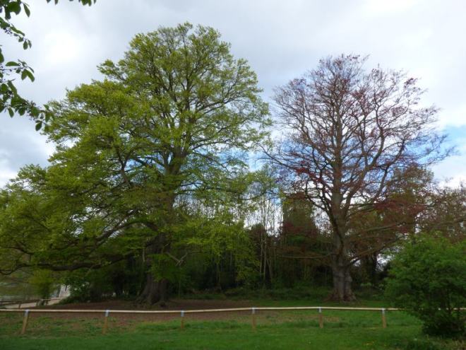
The heart rotters

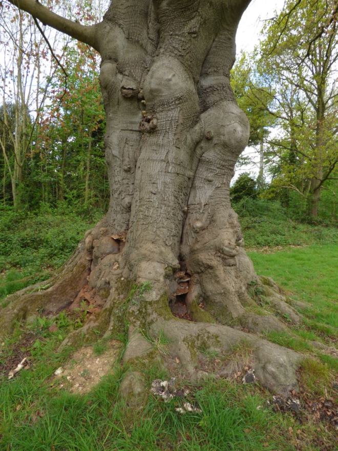
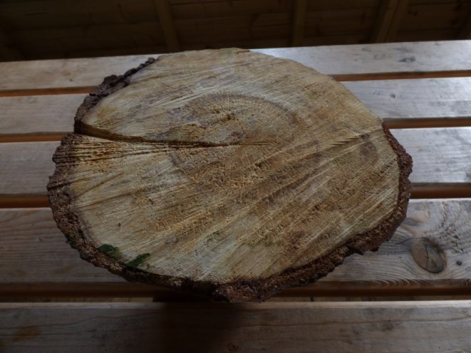
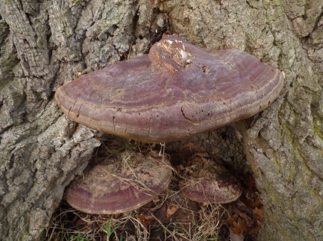
Now, in accepting this, we come on to perhaps the broadest cohort of wood-decay fungi in living and standing trees (fallen trees have their dysfunctional sapwood metabolised like ravens engulfing a fresh meal) we know of: the heart rotters. These fungi will enter the central wood (i.e. ‘the heart’) through exposed areas of this central column by either sufficiently deep root, stem or branch injury – ultimately, there generally needs to be a continuity of viable ‘heart’ substrate, if any significant degree of colonisation is to take place. Where continuity doesn’t exist, or the fungus finds itself limited to only a certain area, the only manner in which is will typically be able to continue existing is by (1) exiting and finding another host or (2) biding its time and waiting for currently functional sapwood to become incorporated into the heartwood or ripewood, in which it can then spread into – assuming the tree doesn’t lay down defensive barriers that cannot be breached, in order to protect its sapwood, which is in itself a pursuit undertaken to safeguard and hence sustain the high-moisture content of its sapwood (contrary to Shigo’s model, which infers compartmentalisation is largely there with the end in mind of prohibiting fungal succession into the wood). When we look at the ripewood of beech (Fagus sylvatica), we can observe this phenomenon very well – a rosy-coloured ripewood (red heart) with lots of separation lines between instances of decay successions. Ganoderma australe, Ganoderma pfeifferi and Ganoderma resinaceum will afford the most acutely observable examples of this, given their ability to breach such zones by metabolising the phenolic deposits laid down by the tree (this touches upon the idea of a fifth wall in the CODIT model, which will be discussed more later on).

laetiporus-taxus-baccata-yew-3
An old Laetiporus sp. on yew (Taxus baccata).

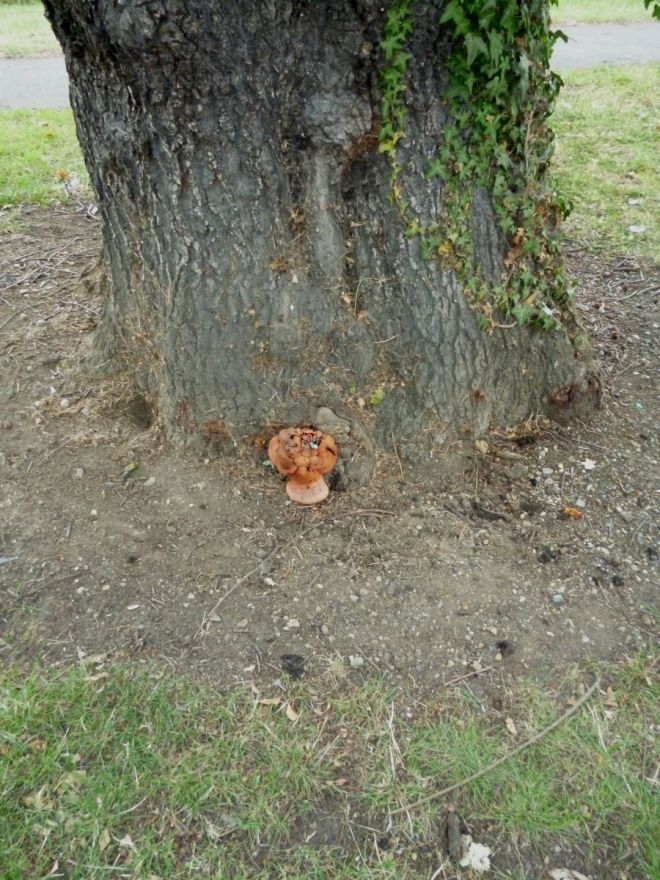
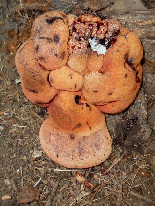
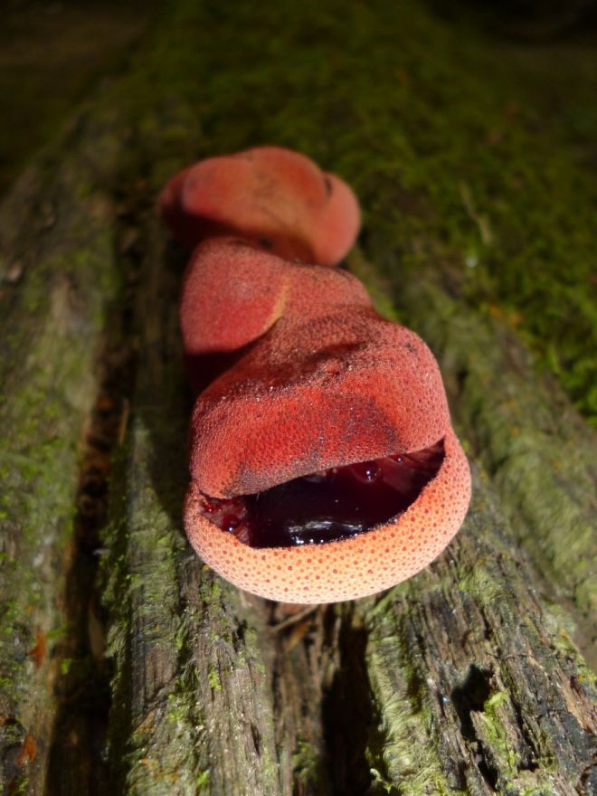
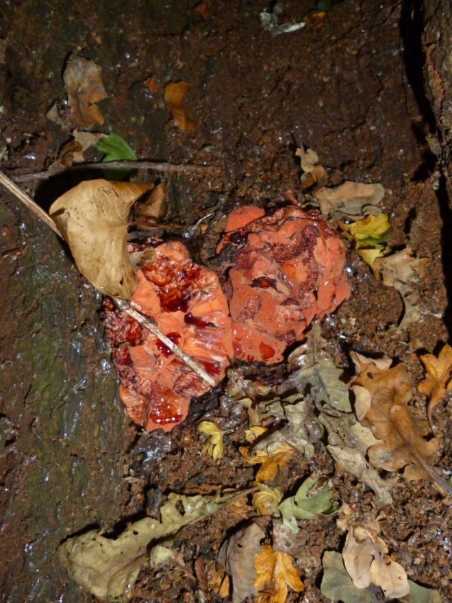
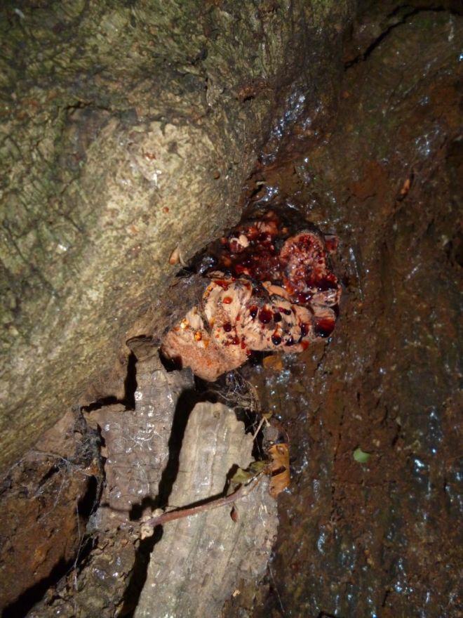
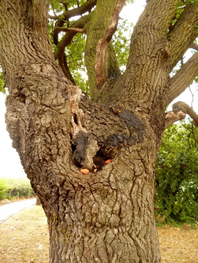
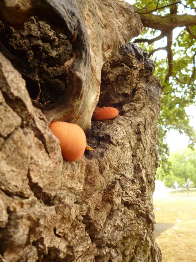
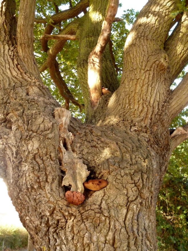
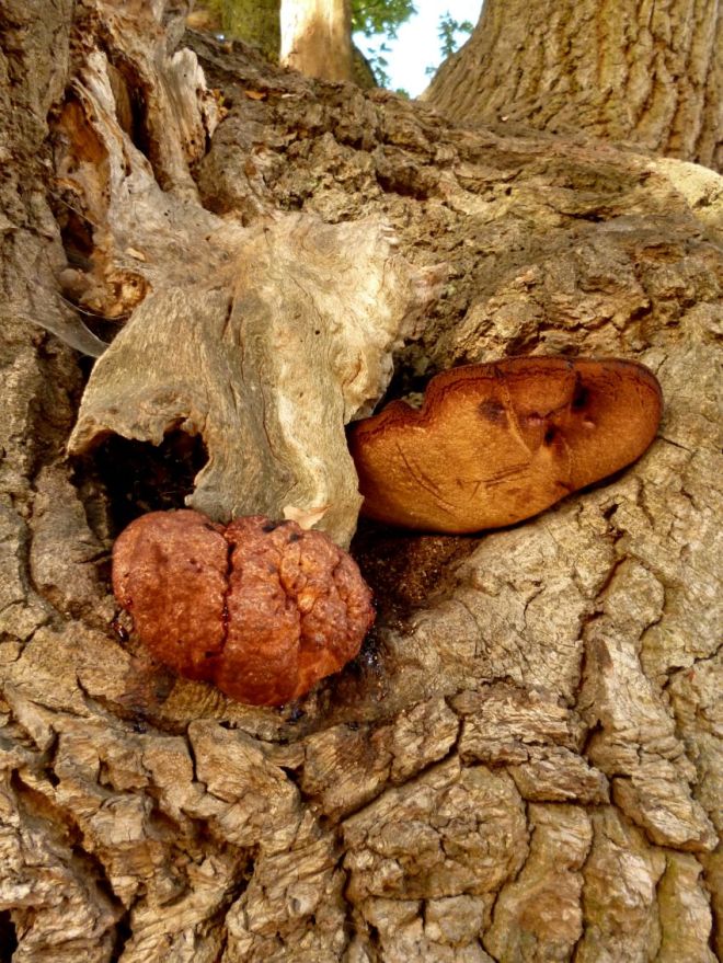
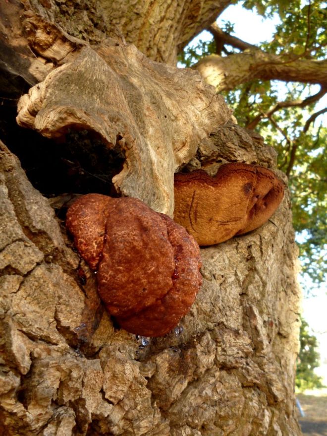
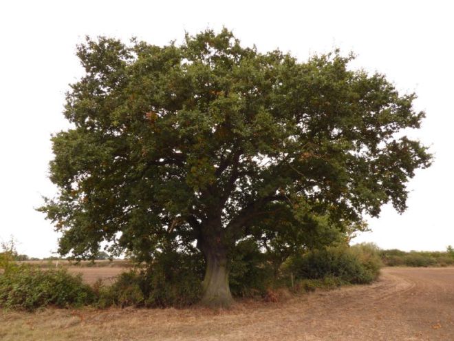
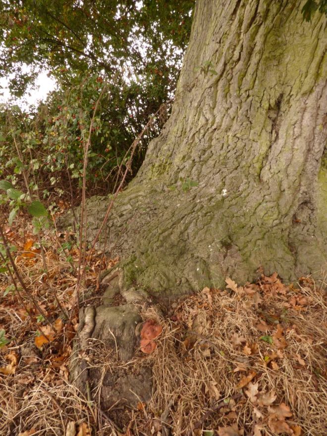
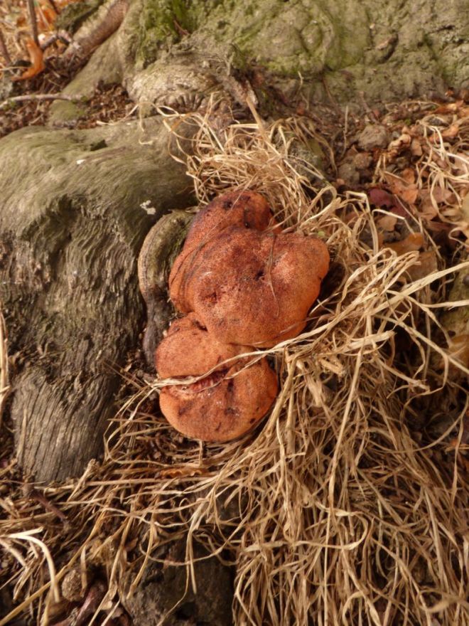
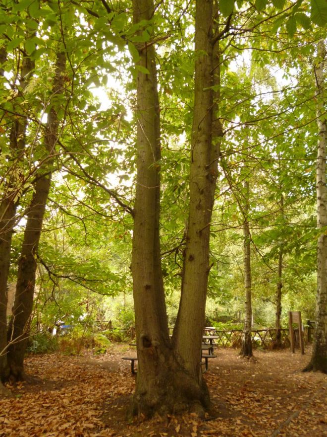
By virtue of the heartwood or ripewood being the least inhospitable (heartwood, in particular, is still often disgustingly harsh, as regards its environment), we can observe immediately some species-specific associations. For instance, the dyer’s mazegill (Phaeolus schweinitzii) will frequent gymnospermous hosts, such as the cedars (Cedrus spp.) and pines (Pinus spp.), whilst chicken of the woods (Laetiporus spp. – often L. sulphureus, though by all means not always, as we will see) will be found often on oak (Quercus spp.), sweet chestnut (Castanea sativa) and yew (Taxus baccata) – it can, indeed, be found on other hosts, as well. On the face value of things, these three tree species have seemingly little in common, though all three have extractive-rich heartwood that this genus can metabolise effectively. In terms of why the genus is referred to and not the exact species, Lynne is of the stance that what we term ‘chicken of the woods’ and default to as L. sulphureus is actually a variety of different species each with their own specialisations – perhaps even down to a specific host tree species (notably for yew, where the chicken species is most pertinently viewed as being a different one).

From a tree management perspective, Lynne then addressed the importance of the heart rotters – what is their impact? Put simply, they change the way in which we view the tree from a safety perspective; as in, when fruiting bodies of heart rotters are identified, if the tree is standing and there exists a target, management considerations are routinely entertained and sometimes the tree is felled. Additionally to this, however, we have other things to appreciate:

  • heart rotters do a great job at recycling nutrients, which can then be re-assimilated by the tree when they are uptaken back through its roots (including adventitious aerial roots) or the mycorrhizal fungi the roots associate with
  • the wood qualities produced by heart rotters are ideal as habitat for saproxylic insects and nesting birds
phlebia-tremellosa-fagus-sylvatica-pollard-2
The heart of this beech has been hollowed-out by decay fungi. In the process, before its failure, what conditions did this decay provide for insects and, crucially, what habitat does it provide now?

Latent colonisers

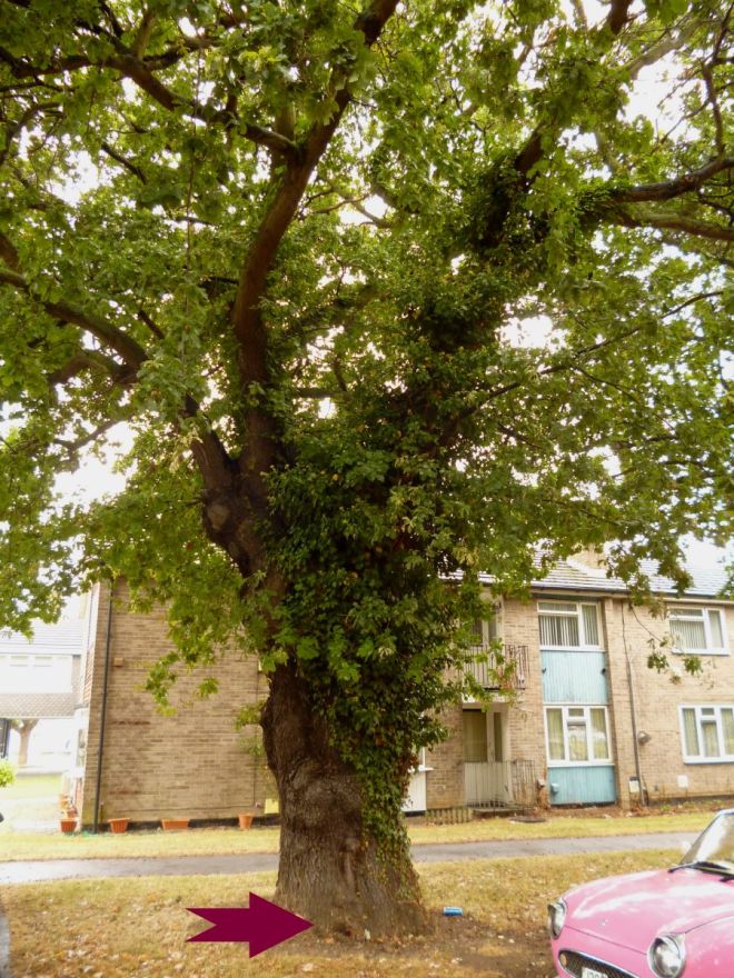
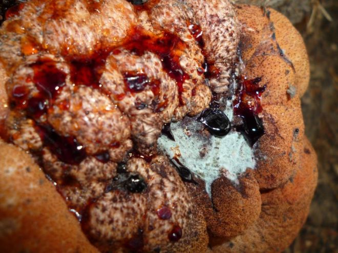
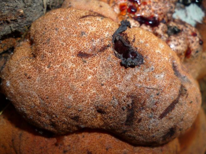
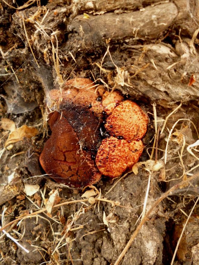
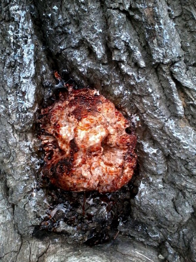
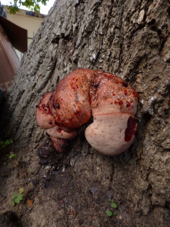
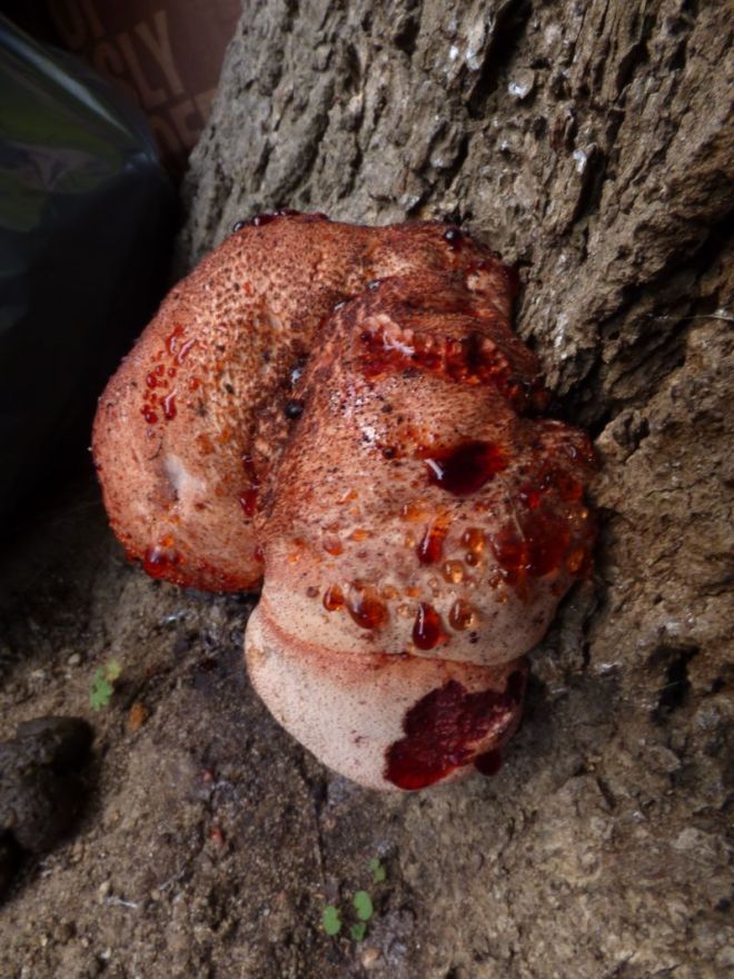
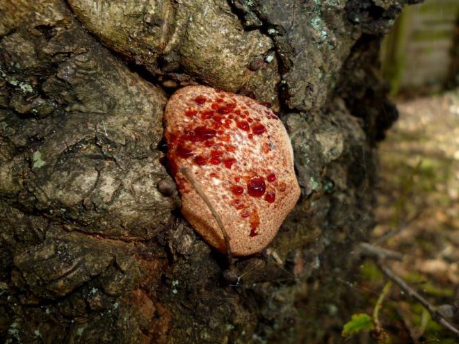
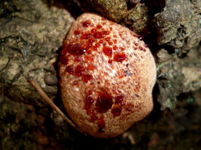
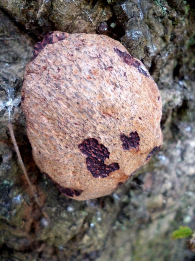
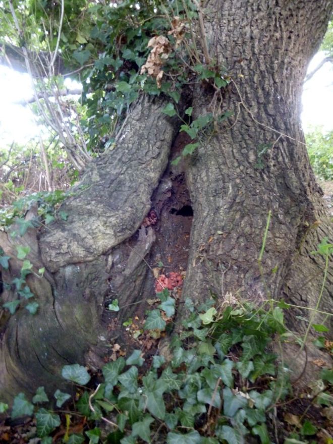
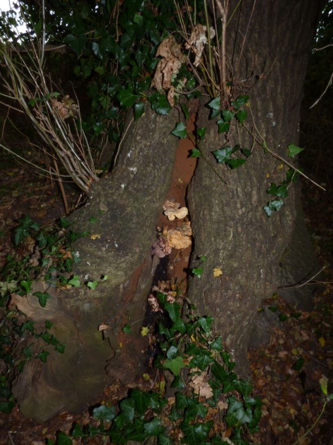
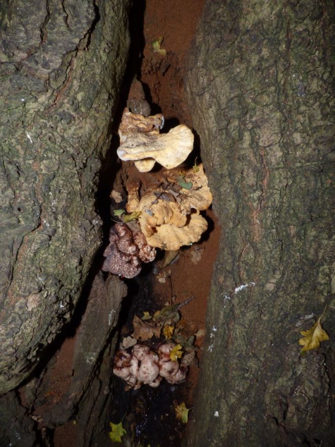
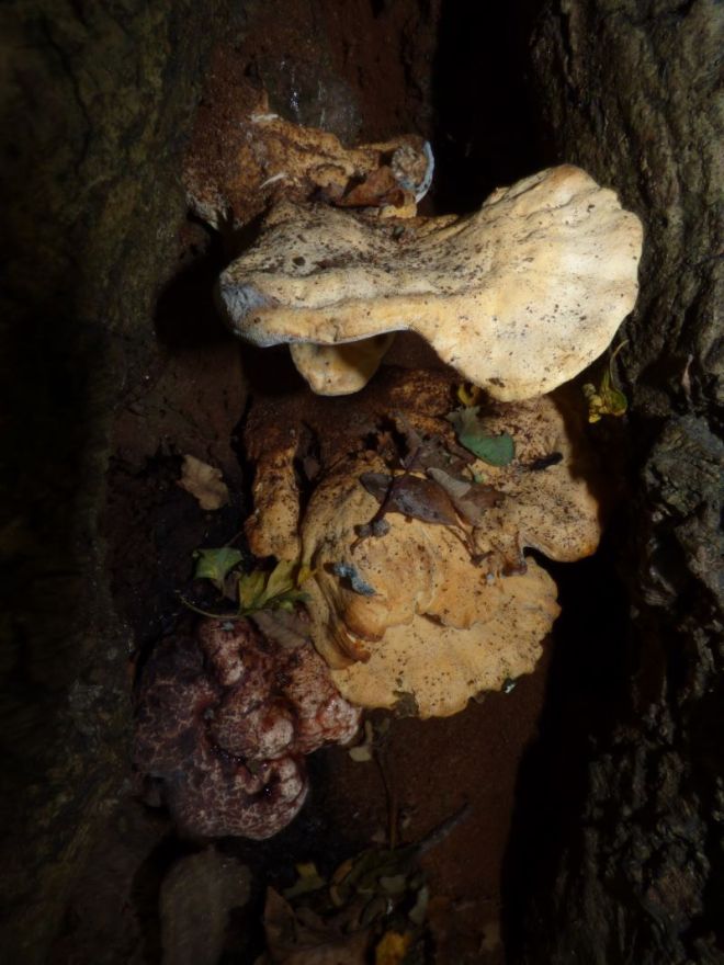
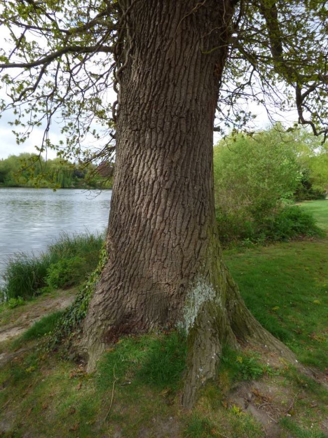
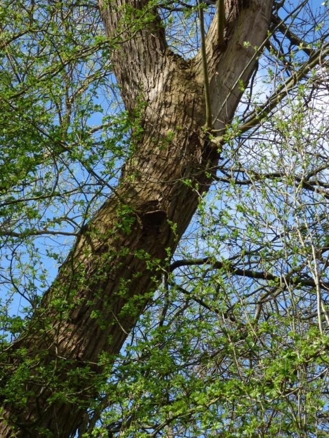
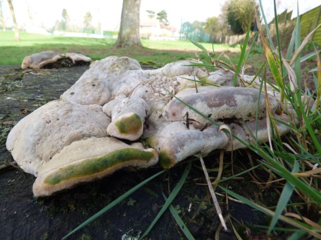
Considered the specialised opportunists, such fungi are present within the sapwood or bark, as propagules (thick-walled resting spores known as chlamydospores). Biding their time until conditions are right, wherein the sapwood becomes dysfunctional through means such as wounding or drought (causing embolism), they are perhaps most acutely observed in the years after drought years where they can trigger the formation of strip cankers and resultant reaction growth by the tree (see the below photo). Thus, this year, in the UK, is one to watch out for, as regards such fungi (it might also explain why Kretzschmaria deusta was so abundant this year, given Ascomycetes love dry conditions, which prevailed last summer).

B nummularia Eutypa spinosa beech canker strip
A strip canker in beech caused by the latent fungus Biscogniauxia nummularia that has induced reaction growth, which can be seen cross-sectionally in the bottom right image).

At this point, a delegate enquired as to whether Massaria disease of plane (Splanchnonema platani), as an Ascomycete, could be prevailing in urban conditions recently, because of dry conditions (such as London, where it was been very severe these past few years but prior to that un-noticed). Discussions continued and Frank Rinn interjected to add his thoughts:

  • massaria progresses quickly in dry conditions
  • recent dry summers have allowed for massaria to thus progress very rapidly (killing branches in as little as three months)
  • the lowering of water tables in cities for the construction of basement levels of buildings has meant that plane trees can no longer tap into groundwater supplies
  • mature plane trees afford the best conditions for massaria; notably lower lateral branches, which are shaded from the rest of the crown and thus may be most prone to stress
  • there have been reports since 1903 from Croatia where large plane trees have shed branches and the massaria fungus was termed the “branch-cleaning fungus”
  • conditions are collectively ideal for massaria to become prevalent, as they stand

Reverting back to latent fungi, Lynne then mentioned that she considers fungi to be latent across a broad variety of trees. For example, the coal fungus (Daldinia concentrica), whilst found most often on ash (Fraxinus excelsior) can be isolated from the sapwood of a great range of different broadleaved tree species in the UK. It is, indeed, only when specific conditions arise that are preferable for this fungus that it begins to create mycelial networks – such conditions might not arise in particular trees, or may arise only after conditions suitable for other fungi have arisen and thus D. concentrica then has no capacity to colonise the substrate. Hence, ash remains the core host of this species, in the current climate. However, for the jelly ear fungus (Auricularia auricula-judae), which is also a latent fungus within the vascular system, having been found largely solely on elder (Sambucus nigra) in the 1950s, it is now found on over 20 host species – this marks a huge increase in host range, prompted perhaps by changing climatic conditions.

Wall V

The CODIT model, offered to us by Shigo, details four walls – as can be seen here. As mentioned by Frank Rinn, Shigo himself was considering the possibility of a fifth wall, though this never ‘made it into’ the model. However, Lynne argues that there is the potential for a fifth one, which hearkens back to what was discussed above, as regards rot within the heart of beech wood).

Specifically, whilst the barrier zone (fourth wall) is a zone laid down at the time of wounding by the vascular cambium, the dynamic responses by the tree that occur in real-time as fungal decay advances constitutes a distinction from this initial barrier. Indeed, as decay advances, living cells within the heartwood or ripewood (they do exist; though through mechanisms not fully appreciated, but thought to be associated with the rays running radially through the wood), in addition to the functional sapwood, will, in order to protect the sapwood and keeps its high-moisture quality intact, will plug woody cells beyond the current zone of decay with extractives and phenolic compounds – this will occur within the sapwood most often, though may also be able to occur in the heartwood around the regions where pockets of living cells exist. This response resultantly produces incredibly dense zones of wood that afford the tree’s sapwood a means of protection, which it would otherwise lack, assuming the barrier zone (Wall IV) failed to contain fungal decay. Of course, if this fifth wall fails, another will form, and so on and so forth.

Ganoderma pfeifferi beeswax beech failure 3
See the myriad of demarcations across this cross-section of a beech that failed from decay by Ganoderma pfeifferi, which suggest a dynamic fifth wall being effective.

Perhaps we will see this idea discussed more in Lynne’s re-write of Fungal Decomposition of Wood.

Fungal succession

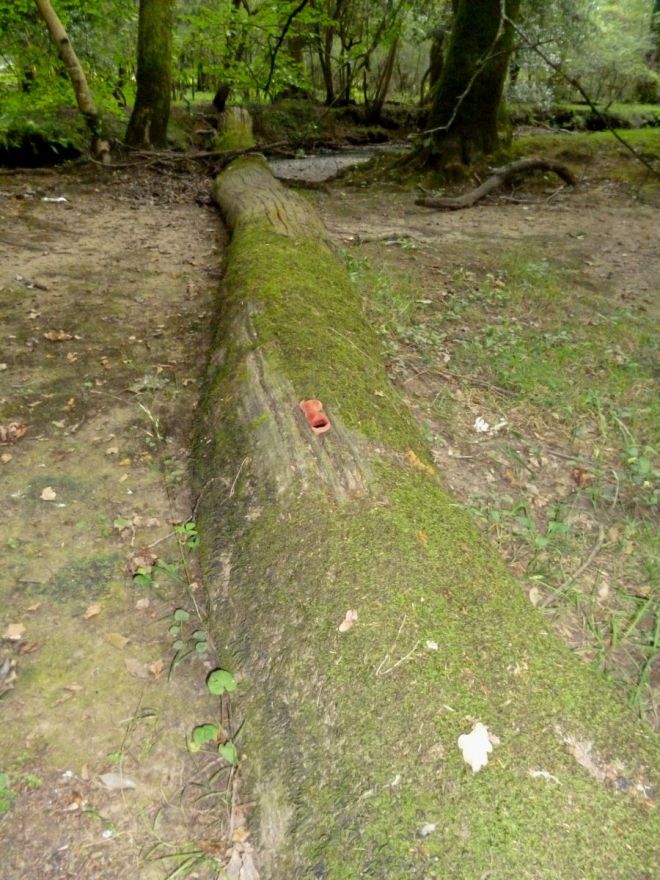
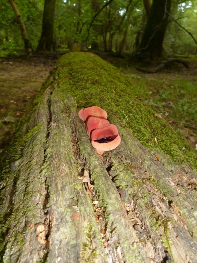
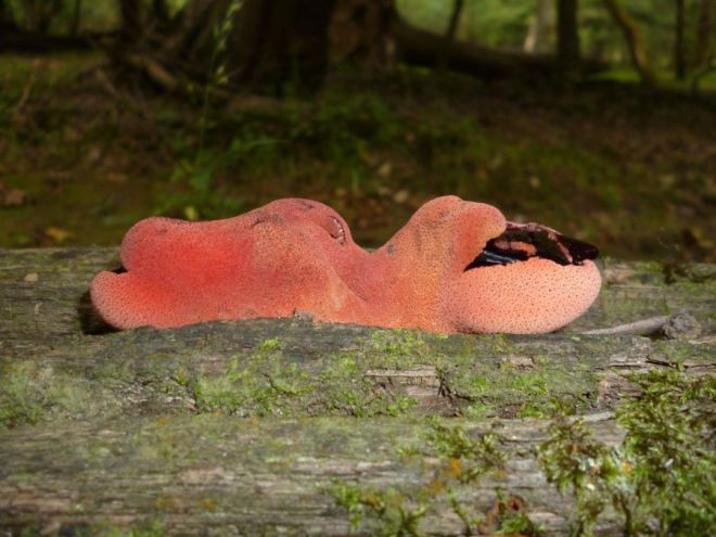
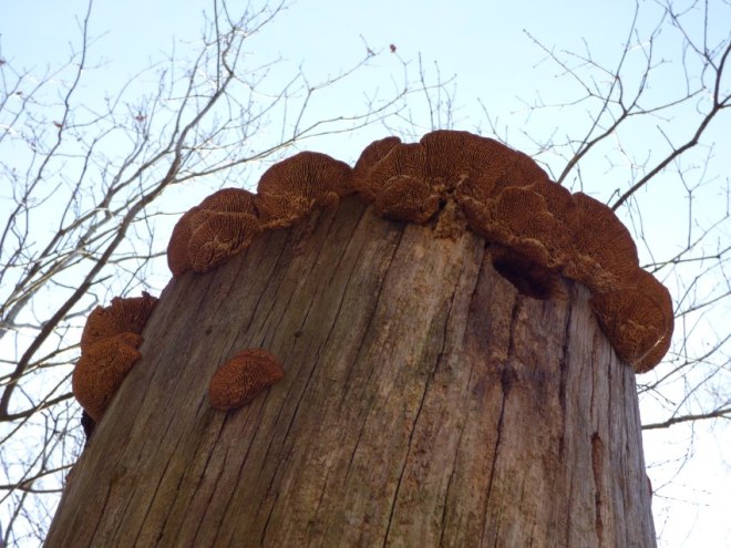
When a tree decays, the fungi that initiated the decay process will not end it. This is because, much like all other ecosystems, as an environment changes those organisms that are best-placed to utilise it change as well. In this sense, fungi are no different – they succeed into a dynamic and ever-altering substrate (wood). Such a phenomenon can be so readily observed when walking into any woodland, when comparing the fungi on standing trees and those in early stages of decay and those much more heavily decomposed. In instances where an entire tree falls and stays largely intact, succession can be most acutely observed, as Lynne detailed with a little help from Ted Green and some research students.

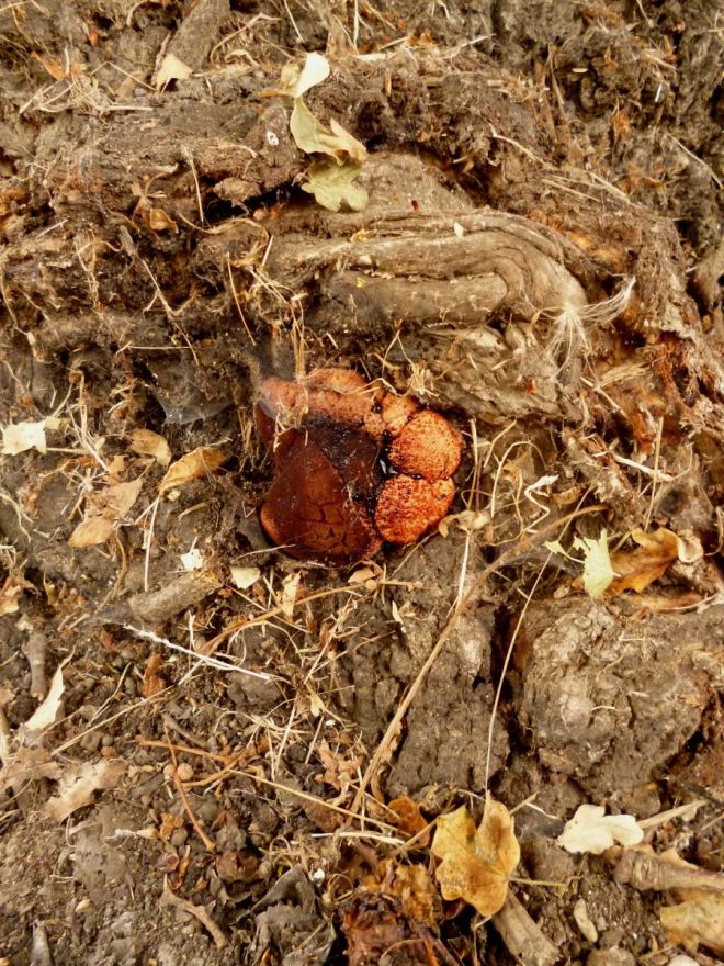
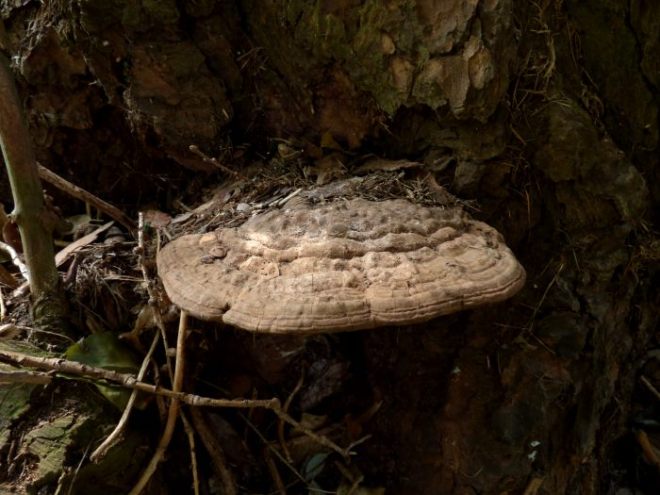
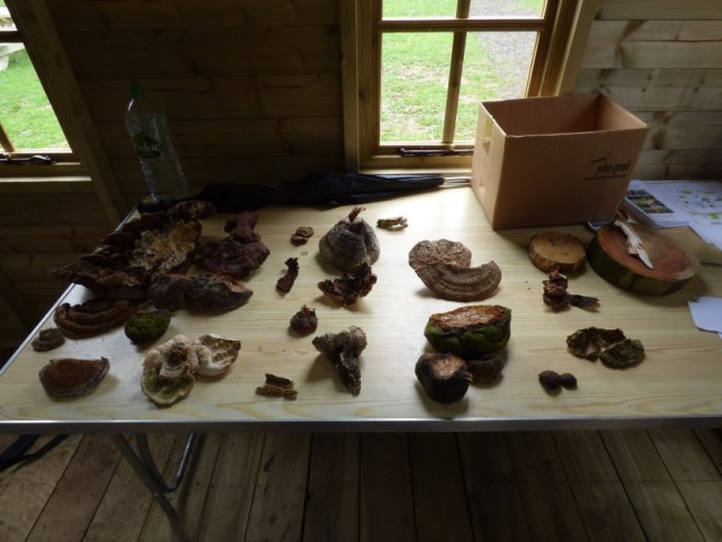
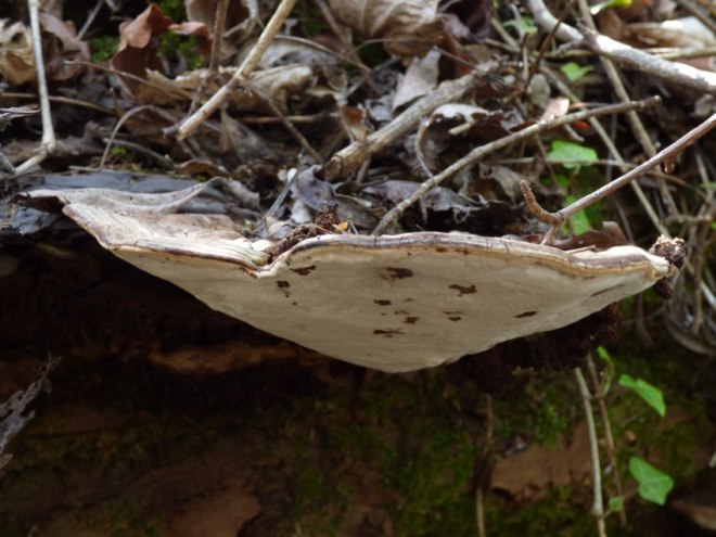
The tree in question, a mature beech, failed and was left, in sections, for ease of its movement into an accessible place, to decay. Along the beech, it was found, through analysis of the wood in the laboratory and by presence of fruiting bodies, different fungi were observed colonising different parts at different stages of decay (as shown below). Such an observation does seem readily apparent, though to have it confirmed through scientific means affords us with an understanding that is more concrete than merely the anecdotal. Indeed, whilst Trametes gibbosa was not isolated from this beech, the presence of Bjerkandera adusta infers that, at some point in the future, T. gibbosa will be found – it parasitises upon the mycelium B. adusta, before then colonising the wood substrate itself. We are, in a sense, therefore, witnessing fungal warfare.

beech fungi succession analysis
The different fungi found at different parts of the beech at different stages of the decay process (open in a new tab to see this in a slightly larger size).

Delving further into the notion of fungal warfare, what is essentially meant is chemical warfare. Fungi synthesise and secrete enzymes, which they use principally to degrade wood, though that can also be used to defend territory or attack other fungi. The result of any fungal battle can be one of four things:

  • deadlock, whereby neither fungi gains any ground against the competing fungus
  • replacement, whereby one fungus loses its territory entirely by the other
  • partial replacement, whereby one fungus loses of some its territory to the other
  • mutual replacement, whereby the fungi essentially ‘trade’ places with one another and neither gains any net ground

So how does one determine the outcome of any such skirmish, you ask? Unfortunately, there are so many variables in play that even pitting two fungi against one another in a laboratory is only going to give a slight allusion to what really occurs, though there does nonetheless exist a limited hierarchy of combativeness from which we can assume who the victor will be, under most circumstances (see here at 25:09 timestamp).

Of course, even in assessing this we still have so many caveats to throw in. For example, where moisture conditions are drier because the wood is more exposed, Ascomycetes (i.e. Hypoxylon spp.) will have a better time in securing more wood substrate, as they operate effectively under dry conditions. Indeed, the wood qualities of the substrate itself will even play a role – did the tree uptake pollutants during its life or is it exposed to such pollutants currently, for example. More crucially, if a dead piece of wood (or entire tree) is standing and has thus been subject to relatively dry and exposed conditions suddenly falls to the woodland floor, those fungi reigning when the tree was standing will likely succumb to wood-decay fungi adapted to higher moisture levels and cooler more stable conditions.

The next part of this series will be a brief one on bacteria in wood, as discussed again by Lynne Boddy. I hope to have that written up in the coming few days.

Fungal succession and wood decay in living trees – a seminar report (Part II)

Fungal succession and wood decay in living trees – a seminar report (Part I)

See part II here.

First and foremost, it is absolutely critical for me to extend my deepest thanks to Jon Hartill of Hartill Trädexpert for organising such a superb single-day event (here is an overview of the itinerary) in Sweden. Indeed, bringing together three such critical speakers (Ted Green, Lynne Boddy and Frank Rinn) who, as was expected, but more acutely so in hindsight, worked well as a unit and offered much in the way of vital information and data, was not likely a simple task. Of course, it worked, and my 12 pages of hand-written scrawlings is testament to this. Thus, the purpose of this blog post (or posts, should I say) is to share the information gathered, so that it can be disseminated more widely and stimulate thoughts amongst the internet audience.

sweden delegates seminar fungi decay trees
Some of the delegates from this seminar. As you’ll see from the below picture, the setting was also rather fitting for a discussion on trees…
sweden delegates seminar fungi decay trees 2
Say hello to the town of Kungalv!

Ancient trees – what secrets remain?

The day was opened by Ted Green, the founder of the Ancient Tree Forum. Here, however, his role was not so much to discuss what is known of our ancient trees, but of what we still need to know – what don’t we know? Granted, there’s probably an utterly frightening amount we are yet to understand, though Ted’s talk was not to wallow in such an angst but instead to prompt directed focus towards aspects of ancient trees we really do need to understand next.

Principally, it was important to set the tone of the presentation: hollowing is an entirely natural process, which very few ancient trees escape. In fact, do they even want to escape such a ‘fate’? Arguably, the answer is no. Anyway, at this early stage, Ted made the distinction between the decay of central wood, separating the decay of heartwood (i.e. Quercus) and the decay of ripewood (i.e. Fagus). ‘True’ heartwood forms when phenols and other extractives and toxic substances to fungi are deposited within the non-living (largely) woody tissues, as sapwood becomes redundant. Ripewood, conversely, forms via different mechanics associated with wounding and other events. However, this was a point posited just to illustrate context, more than anyway.

The real ‘meat’ began when Ted began assessing where the notion of hollowing being bad came from: forestry. As an economic practice, it is obvious that hollowing would be seen as destructive, as basal decay hampers return upon the investment of a stand. Despite this, when moving away from forestry, we can observe that hollow trees don’t drop like flies under wind-loading events – as was the case when the 1987 storm (hurricane) hit the UK and hollow trees stood whilst solid neighbours fell. Certainly, hollow trees did fail and when they did it was oft at the point where the internal hollow met sound wood, though the general jist is that hollowing does not necessarily infer a risk of failure beyond that of what a solid tree would be considered to possess. Ted speculated that this hollowing meant that, under wind loads, the main stem could flex and ‘safely’ deform (a bit like a hosepipe when squeased slightly) under tension and torsion, thereby protecting the stem from forces that could overload a solid stem, which cannot deal with such a load in a similar manner (because it is not hollow, to any degree). In oak, for instance, Pseudoinonotus dryadeus (the Eiffel Tower fungus) can actually be seen as a fungus that aids with tree stability, by prompting pronounced buttressing and the creation of wood that over-compensates for the more centralised decay (as was discussed by Frank Rinn later on), whilst also allowing for the oak to deal with wind loading more effectively, given the presence of the hollowing internal to the trunk.

Pseudoinonotus dryadeus colonisation senescent old 7
Strong buttressing as caused by Pseudoinonotus dryadeus decay on oak. Does the internal hollowing even impact upon the tree’s structural stability, assuming the buttress roots laid down afford the necessary support? Thus, dooes pruning even have a beneficial impact upon reducing risk, when accepting that pruning damages the tree’s ability to photosynthesise and thus manufacture the sugars demanded for wood formation in these buttress zones?

Indeed, reaction growth comes in forms beyond buttressing – stems also flute; sometimes, quite majorly, in mature and veteran trees. This fluting can, in times of wind loading, afford the tree additional stability, through the over-compensated high wood quality, which allows the tree to deal with loading forces by acting akin to a coiled rope. Certainly, torsional loading against the direction of fluting is going to be a marked issue, though otherwise such fluting can be beneficial for stability – even when there are appreciable central hollows. In fact, this led on to another important point: the extent of hollowing and the resultant residual wall thickness (think Mattheck’s t/R or part of the Wessolly’s Statics Integrated Approach model) means very little, as it assesses a tree in blatant disregard for its wider context (exposure, lean, leaf area, wind drag coefficient, management history, the off-set nature of the hollow, etc). Without appreciating these factors, of which there are numerous, how can one state that hollowing is bad and will increase the risk of tree failure?

The talk by Ted then actually moved away from hollowing somewhat and onto other aspects of ancient trees. Specifically, the practice of pruning arose, wherein Ted commented that pruning back to the branch collar in older trees is potentially more destructive than leaving stubs – stubs that will afford epicormic growth and the formation of a new crown / part of the wider crown area. Interestingly, he used the example of beavers felling trees not at their base but a little up from the base, from which these trees oft repsrouted and formed natural coppice. The same, he suspected, could be the case for aerial pruning – leave a stub.

This consideration took Ted onto further points, of which the main one was that of where trees fail most routinely. Indeed, aroung 70% of all failures within a tree at at the branch level – of these, many fail not at the collar but out along the branch itself, thereby leaving a stub. Using examples of trees from Windsor and elsewhere that failed in such a way, he demonstrated that new but lower crowns were formed in the years after; even in cases where every single major limb failed on the tree, thereby creating a tree form like what can be seen in shredded trees.

veteran tree natural shred wind hurricane
An oak tree that lost almost all of its crown in the years after the 1987 storm. Only one limb actually remains (red square). The other limbs all failed (green arrows) and have since resprouted and formed a new crown. Should we be managing our older trees like this and, to extend this question, should we be managing all of our trees like this, if we are to mimic natural forms of failure?

Further to this, Ted got talking root failure in strong winds. In his experience, following the 1987 storm, some older trees began to decline either partially (in select area of the crown) or wholly in the years following, for no outward apparent reason. Ted’s suspicion is that, in place of aerial failure, structural roots failed and the connectivity to specific connected parts of the crown from these roots thus was severed, triggering localised dieback and retrenchment. Therefore, the question of whether localised root plate damage caused localised aerial dieback in older trees was asked and, assuming the answer was that this does occur, it could actually be beneficial for the longevity of the tree – it can retrench, form a lower crown and thus increase its safety factor. In fact, it would also suggest that the occurrence of fungi such as Meripilus giganteus on older trees, in certain instances, would be as a consequence of saprotrophism, in place or parasitism – the fungus follows the root damage and metabolises the severed roots. I then asked Ted whether he thought that the same phenomenon could occur in grazing ecosystems, in which cows, pigs, sheep or otherwise are grazed amongst wood pasture. His answer was one of grazing also being a cause of retrenchment, wherein grazing pressure damages certain structural roots and this leads to subsequent localised aerial retrenchment.

root damage crown retrenchment tree wind
Can select root damage under wind-loading conditions bring about select crown retrenchment, through the connection of certain roots to certain portions of the crown? If so, can this damage, assuming not all roots are severed, improve the longevity of the tree and increase its stem’s safety factor, in spite of hollowing and major yet probably transient dieback?

Other questions raised within Ted’s talk were as follows: (1) are animal pharmaceuticals, often in the form of de-wormers, harmful to the mycorrhizal networks and the tree’s rhizosphere (i.e. soil biota), when they are excreted by the animal in the vicinity of the tree?, (2) is acid rain the most damaging impact upon our old trees, because of its impact upon the soil?, (3) are earthworms the most crucial soil organism for older trees, given their ability to aerate soil and to recycle nutrients by consuming absiced leaves (including those where over-winter pathogens reside, such as oak mildew), and (4) does stress throughout the tree’s life give it the best chance of reaching the veteran or ancient stage, when noting that slower growth and a more responsible management of energy is more sustainable? To these questions, we do need more research.

Take-away points from this talk are, therefore:

  • major hollowing of trees isn’t perhaps an inherently bad thing – notably in older trees,
  • we need to understand the species-specific and age-specific impacts of fungal decay upon trees before confidently exclaiming an increased and unallowable risk of failure,
  • a tree’s situation and history has a direct and marked impact upon the risk brought about by a hollow
  • damage to tree roots on older trees and the possible associated crown effects demands more investigations,
  • we need to determine how we should be pruning older trees, if we are concerned with their longevity, and
  • the rhizosphere’s importance for the health of older trees is a very viable area of research

I’ll write part II up in the coming times, which will focus on Lynne Boddy’s presentations. The third part will be Frank Rinn’s incredible afternoon talk.

Fungal succession and wood decay in living trees – a seminar report (Part I)

Fistulina hepatica’s anamorphic version: Confistulina

Having been informed earlier by a friend that they have an article coming out soon in the journal Field Mycology and having then read the article, I considered it important to build on the information and photos shared in the manner I am most familiar: ramblings. I do not want to spoil the article so please do source the article yourself, though as a form of executive summary, this fungus, Confistulina hepatica, is the anamorphic (asexual) stage of the fungus Fistulina hepatica (beefsteak), which is common on oak and sweet chestnut. I have found it once on beech, though it generally sticks to the first two hosts. This anamorphic stage produces asexual spore, which adorn the fruiting body’s outer portion in abundance. The reason for its emergence is not known, though it might be weather-related. More information is absolutely required from across its host range – not just England!

Disclaimer: The older finds, because I didn’t know what I was looking at, are quite poor photographs. A huge shame, but alas!

Disclaimer 2: I suspect many of these are the anamorphic stage Confistulina hepatica, though none were confirmed so please do not assume they all are. This blog post is simply to begin building a knowledge base that builds of the limited resource pool of present.

The first oak that I’ll share is perhaps one of the more interesting of the bunch because, two years in a row (2015 and 2016), the outwardly anamorphic fruiting body appeared in the same location; albeit, in 2015, the fruiting body was larger. Nonetheless, it infers that, in a crude manner, the same mycelial colony has done the same thing two years in succession. Unfortunately, during 2015, I didn’t get a shot of the whole tree so the location of the arrow points to the 2016 occurrence. However, from the positioning of the ivy, we can see the location is the same.

Confistulina hepatica Fistulina anamorphic oak Quercus 1
The arrow points to the location, which is on the southern side of the tree right at the base.
Confistulina hepatica Fistulina anamorphic oak Quercus 2
The 2015 version in its rotten glory!
Confistulina hepatica Fistulina anamorphic oak Quercus 3
The fruiting body sports what looks like burns, in addition to exuding a tar-coloured liquid alongside the more routinely observed reddish exudations common to young fruiting bodies.
Confistulina hepatica Fistulina anamorphic oak Quercus 4
A closer look at the darker liquid from the 2015 version.
Confistulina hepatica Fistulina anamorphic oak Quercus 5
And a puncture wound (so it appears) in the fruiting body.
Confistulina hepatica Fistulina anamorphic oak Quercus 6
And the smaller 2016 emergence!
Confistulina hepatica Fistulina anamorphic oak Quercus 7
Again, we can see the darker liquid exudations. The morphology is also somewhat similar.
Confistulina hepatica Fistulina anamorphic oak Quercus 8
Closer still on the 2016 find.

The next series of shots were taken on my mobile phone back in 2015 and again from oak. Once more, the adornment of the fruiting body with exudations, of which some are darker, can be observed. 2016 saw the fungus return, with a vengeance, and in a different position, again, an anamorphic fruiting body.

Confistulina hepatica Fistulina anamorphic oak Quercus 9
Yes, this oak has been hammered. Poor tree! (2015)
Confistulina hepatica Fistulina anamorphic oak Quercus 10
At the base we can see the fruiting body.
Confistulina hepatica Fistulina anamorphic oak Quercus 11
Oozing everywhere!
Confistulina hepatica Fistulina anamorphic oak Quercus 12
Looks like something out of Alien, quite honestly!
Confistulina hepatica Fistulina anamorphic oak Quercus 34
2016 checking in. What a load of rubbish!
Confistulina hepatica Fistulina anamorphic oak Quercus 35
Further round the tree (note that the 2015 location was to the right) sits an anamorphic fruiting body.
Confistulina hepatica Fistulina anamorphic oak Quercus 36
A close(ish) look.
Confistulina hepatica Fistulina anamorphic oak Quercus 37
Closer still.
Confistulina hepatica Fistulina anamorphic oak Quercus 38
And closer yet further.

From here, we go to another 2015 find. This time, the fruiting body was a little elevated on the oak and evidently emanating from an area of burring / accumulation of dormant buds beneath the bark surface. This one quickly became senescent after perhaps a week so whether it’s Confistulina or not is tough to say – I include photos of both times I visited it. It did not reappear in 2016.

Confistulina hepatica Fistulina anamorphic oak Quercus 13
The first visit to this fungus, which was very small – maybe 5cm across.
Confistulina hepatica Fistulina anamorphic oak Quercus 14
A closer look.
Confistulina hepatica Fistulina anamorphic oak Quercus 15
A week later it had senesced.

The remainder of the photos take us into 2016 and most (but not all!) are taken with a better camera, which is good! First, we venture down to the New Forest and look at a well-decayed oak log, upon which a cluster of fruiting bodies sit atop the log.

Confistulina hepatica Fistulina anamorphic oak Quercus 16
Yes, yes, this photo was taken with a potato.
Confistulina hepatica Fistulina anamorphic oak Quercus 17
A very odd form but once again we can see the tarry liquid.
Confistulina hepatica Fistulina anamorphic oak Quercus 18
A side profile. Looks like a toe!
Confistulina hepatica Fistulina anamorphic oak Quercus 19
Yep, definitely a toe – a wrangled one, at that.

The next series are taken a month apart (late August and late September). What’s curious with this one is that we can see two anamorphic fruiting bodies and, come September, Laetiporus sulphureus fruiting directly alongside. The host, as can be seen, is oak, which is in a hedgerow of oak and ash.

Confistulina hepatica Fistulina anamorphic oak Quercus 20
Fading light caused the blur!
Confistulina hepatica Fistulina anamorphic oak Quercus 21
At the base we can see a dueo of fruiting bodies.
Confistulina hepatica Fistulina anamorphic oak Quercus 22
The first…
Confistulina hepatica Fistulina anamorphic oak Quercus 23
…and the gruesome second.
Confistulina hepatica Fistulina anamorphic oak Quercus 25
A month later a wild chicken appeared!
Confistulina hepatica Fistulina anamorphic oak Quercus 26
Two, in fact. Here we see the beefsteak and chicken side-by-side.
Confistulina hepatica Fistulina anamorphic oak Quercus 27
And here it’s in the distant murk.

Now, we move to a curious case of two fruiting bodies next to one another, again on oak, where one became a teleomorph and the other an anamorph. Why? Who knows. Very interesting, however, as I am sure you can appreciate.

Confistulina hepatica Fistulina anamorphic oak Quercus 28
An oak pollard by a swing. Look at the old pollard head.
Confistulina hepatica Fistulina anamorphic oak Quercus 29
A duo forming.
Confistulina hepatica Fistulina anamorphic oak Quercus 30
Looking pretty similar. Now watch…
Confistulina hepatica Fistulina anamorphic oak Quercus 31
…they change! No longer are they twins.
Confistulina hepatica Fistulina anamorphic oak Quercus 32
Exactly why this happened is a mystery though I’d really like to know – anamorph left) and teleomorph (right).
Confistulina hepatica Fistulina anamorphic oak Quercus 33
A side profile for comparison with the earlier one taken from the same position.

In October of 2016, I came across this majestic oak in a field. On a buttress root was what appeared to be an anamorphic fruiting body of Fistulina hepatica, which we can observe below.

Confistulina hepatica Fistulina anamorphic oak Quercus 39
Yup – majestic.
Confistulina hepatica Fistulina anamorphic oak Quercus 40
Also majestic??? (not sure!!)
Confistulina hepatica Fistulina anamorphic oak Quercus 41
Eh, nope. A bit more ugly, to be fair. Has the outward character of an anamorph though please don’t assume it is (wasn’t confirmed via microscopy).

Lastly, here’s one I suspect on sweet chestnut. I didn’t return to check how it did, though this year I’ll certainly keep more of an eye out and be more thorough in my inspections of the samples! Please share any examples you might have found of this, too – it’s important we build up a knowledge base.

Confistulina hepatica Fistulina anamorphic sweet chestnut Castanea 1
A lovely old coppice stool.
Confistulina hepatica Fistulina anamorphic sweet chestnut Castanea 2
Here sits the sample.
Confistulina hepatica Fistulina anamorphic sweet chestnut Castanea 3
Whether this is or is not is tough to say, though it does resemble an anamorphic stage somewhat.
Fistulina hepatica’s anamorphic version: Confistulina

Interesting cases of wood-decay fungi

I have been absurdly busy so haven’t been blessed with the time to get some blogging in, though I have been graced with thirty minutes of time this evening so without any further utterings we’ll delve right into the good stuff – trees and fungi (in my usual frenetic and incoherent manner). Plus, listening to some early Hawkwind has really got me in the mood to do something useful!

The cases are all from an absolutely splendid park down in Maidstone – Mote Park. Honestly, if you live anywhere near there, do pay it a visit and explore as much as you can (it’s massive!). The sheer abundance of mature and veteran trees provides for a magnificent display of fungi and, so I am told, there is a need to record the saproxylic insects on site on the many monoliths and moribund trees.

To kick this post off, I take you to a very interesting case of Pseudoinonotus dryadeus on oak – three of them, all of which are within 8-10m of one another and share a crown. All fruitings of the fungus are historic though its presence on all three trees makes for some tempting considerations – namely, the synchronicity of fruiting (are they similar genotypes?) and the means of colonisation (spore or something else?). Indeed, I can only infer some sort of fungal mysticism or sorcery in positing both aspects for consideration (there is no proof of either, per se), though it did make me think. Perhaps it will make you readers think as well! (!?)

Pseudoinonotus dryadeus colonisation senescent old 1
The recipients of the Manchurian Candidate!
Pseudoinonotus dryadeus colonisation senescent old 2
Exhibit one!
Pseudoinonotus dryadeus colonisation senescent old 3
At the base (to the left).
Pseudoinonotus dryadeus colonisation senescent old 4
Quite an old bracket but a bracket nonetheless. Lovely buttressing, too.
Pseudoinonotus dryadeus colonisation senescent old 5
Exhibit two. More brackets and more buttressing.
Pseudoinonotus dryadeus colonisation senescent old 6
Shame these got yanked off, too.
Pseudoinonotus dryadeus colonisation senescent old 7
Exhibit three! SOme glorious buttressing here, yet again. Thus the fungus gets its common name: the Eiffel Tower fungus.
Pseudoinonotus dryadeus colonisation senescent old 8
Old, dead (not really but who cares for semantecs?) but not forgotten.

And then…and then…more Pseudoinonotus dryadeus – literally 100 yards down the same path. Oh how Mote Park delivers! This example also really does demonstrate the magnificent buttressing induced by its decay on oak, as you’ll see.

Pseudoinonotus dryadeus mature oak butt decay 1Pseudoinonotus dryadeus mature oak butt decay 2Pseudoinonotus dryadeus mature oak butt decay 3Pseudoinonotus dryadeus mature oak butt decay 4Pseudoinonotus dryadeus mature oak butt decay 5

Would you then believe it? Essentially opposite (no joke) were two colossal beech trees fenced-off (as if that ever stopped me??!) that, as anyone who has seen mature or veteran beech buttressing all over the place like egg whites pour out of a broken egg when broken too aggressively (nice analogy? – likely not), drew me in. Was I disappointed? Not at all! Ganoderma australe and Meripilus giganteus all over the option.

Fagus sylvatica mature buttressing Ganoderma Meripilus 1
Good cop (right) bad cop (left) – something something pun something something copper beech and tell better jokes
Fagus sylvatica mature buttressing Ganoderma Meripilus 2
The copper beech to the right with roots all over the option.
Fagus sylvatica mature buttressing Ganoderma Meripilus 3
Ganoderma australe and Meripilus giganteus – dual decay. Decay squared? That raises a good thought – is decay by more than one fungus simply a cumulative issue or instead a geometric or even negatory issue (i.e. is decay of two types ‘less serious’ than from one only)? Question galore and no answer. Someone ask an expert!
Fagus sylvatica mature buttressing Ganoderma Meripilus 4
Merip (foreground) and Gano (background). Also plenty of blades of grass for the monocot enthusiasts who stumbled across this blog because I wrote the word monocot.
Fagus sylvatica mature buttressing Ganoderma Meripilus 5
Ganodeerma australe being illuminated by a sunburst.
Fagus sylvatica mature buttressing Ganoderma Meripilus 6
The diddy little brackets further up the stem weren’t blessed with sun.
Fagus sylvatica mature buttressing Ganoderma Meripilus 7
And between some more buttresses there were some more Ganodermas.
Fagus sylvatica mature buttressing Ganoderma Meripilus 8
Onto the plain old beech now. Exquisite buttressing here!
Fagus sylvatica mature buttressing Ganoderma Meripilus 9
Did someone say fungi?
Fagus sylvatica mature buttressing Ganoderma Meripilus 10
Nope – nothing to see here.
Fagus sylvatica mature buttressing Ganoderma Meripilus 11
Nor is there anything to see here. I’m just tired of writing captions!
Fagus sylvatica mature buttressing Ganoderma Meripilus 12
Oh look, finally. Something. Some nice husks. Probably some Ascomycetes on them (Xylaria carpophila).

To finish up, because I’m getting tired and I am up early tomorrow, here’s something to sit on whilst you ponder the plethora of ultimate questions spewed forth from my mind with little restraint – a dryad saddle. The host? Not sure – lots of ash about though one can never rule out sycamore (unless you’re in the middle of a Douglas fir plantation?). These had actually already over-matured, which means you can see dryad saddle (i.e Cerioporus squamosus – named, prior to that, Polyporus squamosus) out there if you look!

Dryad saddle Polyporus Cerioporus squamosus 1Dryad saddle Polyporus Cerioporus squamosus 2Dryad saddle Polyporus Cerioporus squamosus 3Dryad saddle Polyporus Cerioporus squamosus 4

Interesting cases of wood-decay fungi

Ganoderma australe on larch (Larix decidua)

An interesting relationship going on here, which has been confirmed as Ganoderma australe colonising a larch specimen through microscopic analysis by a professional mycologist. This particular larch is wonderfully clad with ivy, though is still alive (just!). I came across this approximately a week ago and was quick to get it assessed, given the rarity of the relationship.

Not much to add here, besides reiterating it being a very infrequent occurrence – if at all recorded before and subsequently confirmed, in the UK. Cool, eh!

Ganoderma australe Larix decidua larch UK 1Ganoderma australe Larix decidua larch UK 2Ganoderma australe Larix decidua larch UK 3Ganoderma australe Larix decidua larch UK 4Ganoderma australe Larix decidua larch UK 5Ganoderma australe Larix decidua larch UK 6Ganoderma australe Larix decidua larch UK 7Ganoderma australe Larix decidua larch UK 8

Ganoderma australe on larch (Larix decidua)

A trip to Aldenham Country Park – trees and fungi

With the weather remaining fair, in spite of the onerous musings spouted from the verbal orifices of the meteorological office, getting out at the weekend to explore new sites is still very much on the cards. Today, a group of us went over to Aldenham Country Park in north-west London, to search for interesting fungi on trees; as if a weekend would yield any other result!

We started the day by doing something socially reprehensible: bringing in fungi collections for display. As the below photos show, my collection is growing in extent, though is dwarfed in literal size by another collection, which essentially involves monster brackets that are, in some cases, still clinging to the very substrate that provided them with their life.

polypore collection fungi
My collection, consisting of fruiting bodies of fungi including the genus Ganoderma (top left), the genus Trametes (bottom left), Fomes fomentarius (top middle), the genus Phellinus (bottom right) and Coriolopsis gallica (bottom right).
polypore collection 2
Another collection, set up almost like a demonstration of the solar system (with the Perenniporia fraxinea on the poplar being the sun, of course!), including Fomes fomentarius (a monster one), Daedalea quercina and, as stated, the Perenniporia fraxinea on the poplar wood.

Before sharing some finds from today, it’s almost important to share some images of more cross-sectional decay as caused by Ganoderma pfeifferi. For those of you with a memory that stretches back beyond a mere seven days, you might recall a recent post I made showing a decay cross-section on a failed beech. Below, we see how the fungus’ activity within a branch stub of a beech has resulted in zonal decay, which is somewhat comparable to the other example shared recently – particularly, with regards to the rosing pattern.

Ganoderma pfeifferi internal decay 1
A tiny Ganoderma pfeifferi within the opening of a branch stub wound on beech.
Ganoderma pfeifferi internal decay 2
The cross-section of decay produced by the fungus.

And so, on with the walk we did, quite early on we wandered past an old poplar stump with some quite extensive Rigidoporus ulmarius decay. Indeed, as is quite routine with this fungus, the internal hollow was clad aplenty with small brackets, whilst the outside sported a much more sizeable fruiting body still in an active phase of its existence. Evidently, a new hymenium has recently been laid down, suggesting that this fungus is soon ready to begin producing spore for the coming season.

Populus Rigidoporus ulmarius stump decay 1
Rigidoporus ulmarius acting as a saprotroph on this senescent stump.
Populus Rigidoporus ulmarius stump decay 2
Quite a nice one, actually! Good morphology.
Populus Rigidoporus ulmarius stump decay 3
Looking inside the hollow, not only can we see that it is used as a bin, but also to house many small fruiting bodies of this fungus.

Very soon after this sighting, a fallen poplar log with Oxyporus populinus was discovered. I admit to only having seen this fungus twice, of which this find was one, so for me this was particularly exciting. In fact, the single fruiting body was rather massive and easily discernible by the quite brilliant tube layers separated by narrow bands of mycelium. Almost directly adjacent to this was a fruiting body of Ganoderma applanatum, as could be determined morphologically by the very thin cuticle atop the bracket (that is crushed easily and cuts very easily) and the extensive damage to the fruiting body, as caused by the yellow flat-footed fly Agathomyia wankowiczii.

Oxyporus populinus Populus log 1
A poplar log hides amongst ivy.
Oxyporus populinus Populus log 2
On one of the cut ends sits this large fruiting body of the fungus Oxyporus populinus.
Oxyporus populinus Populus log 3
The demarcations between each growth spurt are incredibly distinct, in this fungus.
Ganoderma applanatum Populus log 1
A fruiting body of Ganoderma applanatum also sat nearby, on the same log.
Agathomyia wankowiczii Ganoderma applanatum Populus log 3
Underneath, we can see the distinct gall structures caused by the yellow flat-footed fly.
Agathomyia wankowiczii Ganoderma applanatum Populus log 2
We can also see the internal damage caused by the fly, as it develops into its adult form and leaves to lay eggs elsewhere. The very thin upper cuticle can also be seen, which is thicker on Ganoderma australe.

Following the sighting of copious amounts of Daedaleopsis confragosa, our attention was then drawn to a rather sorry-looking beech tree over a well-used footpath. Upon close inspection, both Kretzschmaria deusta and the rhizomorphs of Armillaria mellea could be found, which certainly puts the longevity of this beech as is into doubt. To be honest, in all likelihood it’ll be monolithed, in order to still provide habitat but with the risk removed.

Kretzschmaria deusta beech Fagus Armillaria 1
It even leans over the footpath!
Kretzschmaria deusta beech Fagus Armillaria 2
Both the anamorphic stage of Kretzschmaria deusta and cambial necrosis caused by Armillaria mellea can be seen, in this image.
Kretzschmaria deusta beech Fagus Armillaria 3
Not looking good for this beech!

Around the proverbial corner (it was more like a ten minute trundle) from this beech stood a massive stump of an old poplar. In its prime, this would have been a tree operating on beast-mode, though is now far more modest in size. However, to make up for its literal demise, it now is host to the fungus Trametes gibbosa, which can be seen around one of the two stems.

Trametes gibbosa Populus stump 1
A fortress of nettles guards this poplar stump.
Trametes gibbosa Populus stump 2
Too bad they can’t defend against a zoom lens and / or walking boots and jeans!
Trametes gibbosa Populus stump 3
Some fresh brackets adorn the opposite side of the stump.
Trametes gibbosa Populus stump 4
Quite pretty, to be honest!

Delightfully, this stump also housed a bird nest, which I found only by pure chance when noticing what looked like chocolate mini-eggs! Tucked away impossibly well within a bark crevice was a small robin’s nest (I think), complete with four eggs. Hopefully, this stump will offer enough privacy to enable the chicks to develop well and not get picked-off by predators.

Erithacus rubecula eggs poplar stump tree 1
The arrow shows where the nest is, as it’d otherwise be impossible to see!
Erithacus rubecula eggs poplar stump tree 2
There were four eggs in this tiny nest. Such a great place for shelter and quite absurd that I came across it!

Once we had come across yet more Daedaleopsis confragosa, which I was busy photographing, a friend spotted a single Sarcoscypha coccinea (scarlet elf cup). Somehow, this is the first time I have seen this fungus and I can understand why it’s such a popular one! An absolute gem.

Sarcoscypha coccinea 1
Cheeky! Hiding away under nettles. Almost doesn’t want to be discovered…
Sarcoscypha coccinea 2
Nature’s very own satellite dish!

And then came something I found very interesting: my first ever sighting of the fungus of willow known as Phellinus igniarius. Upon what was either a crack willow or white willow, a few fruiting bodies had grown and the decay had since led to failure of an upper limb, which has since been cut up and left on the ground. The resulting abundance of fruiting bodies on both the tree and sawn logs is a testamenrt to the extensive colonisation of this fungus within the host. The largest bracket, which was a casulaty of the failure, in fact did not senesce and instead reiterated its growth so that the hymenium and tube layer re-grew at an angle perfectly parallel with the ground (known as geotropism / gravitropsim).

Phellinus igniarius Salix alba fragilis sp decay 1
A willow not unlike any other willow – battered by the elements.
Phellinus igniarius Salix alba fragilis sp decay 2
Oh but wait – a fungus! Surely it’s a Ganoderma…
Phellinus igniarius Salix alba fragilis sp decay 3
…nope!
Phellinus igniarius Salix alba fragilis sp decay 4
As we shall see by what is on the floor, upon these logs…
Phellinus igniarius Salix alba fragilis sp decay 5
…Phellinus igniarius! Surprise! (assuming you didn’t read the text and look only at the pictures)
Phellinus igniarius Salix alba fragilis sp decay 6
Quite a significant number of new sporophores are forming, following the fragmentation of this limb.
Phellinus igniarius Salix alba fragilis sp decay 7
Around an old branch tear sits a single fruiting body, however.
Phellinus igniarius Salix alba fragilis sp decay 8
Not unlike a young Fomes fomentarius, really!
Phellinus igniarius Salix alba fragilis sp decay 9
And the main bracket has not perished!
Phellinus igniarius Salix alba fragilis sp decay 10
Using flash photography (literally), we can see the white spore print beneath the reiterated growth, following the change in orientation of this bracket.

To round off, I share a diabolically grotesque example of Ganoderma resinaceum upon Turkey oak. Enough to challenge the gargoyle statues of various catacombs (in both video games and real life, if there exist any!) for the prize of what’s the most vile in appearance, and we’re not talking about the Turkey oak here, this fungus is clearly a shadow of its former self. Nonetheless, it is important we can still identify them in such aberrant form, if we are to appropriate diagnose issues and enact management regimes. Thus, as a sort of encore, I present to you…

Ganoderma resinaceum Quercus cerris weird 1
Nice enough tree, eh!
Ganoderma resinaceum Quercus cerris weird 2
But what is that at the base!?
Ganoderma resinaceum Quercus cerris weird 3
Uhh………??
Ganoderma resinaceum Quercus cerris weird 4
Yeah; uhhh…….?
Ganoderma resinaceum Quercus cerris weird 5
Ganoderma resinaceum!
A trip to Aldenham Country Park – trees and fungi

Burnham Beeches – old trees, wood decay and sun

A huge thanks to Burnham Beeches yesterday for hosting some of us for the day and showing us around the site. Below are some of the stand-outs from the day, which I am certain you will all appreciate!

The importance of functional units

As we can see in the below few images of a particularly striking beech pollard, very little of the structure of the tree needs to remain for the tree to persist as a living and functional organism. In this example, only one unit of vascularity supports a very small crown, though the beech is generally without significant fault. It could, potentially, persist in this state for many decades! Certainly, the two natural ‘props’ that support the crowd through a sort of tripod could, in their eventual failure, be the demise of this tree; assuming the functional unit cannot itself adequately support the crown. Depending on the rate of decay of this two ‘props’, this last vascular strip might (if decay is slow) – or might not (if the ‘props’ fail sporadically) – be able to lay down the necessary wood fibres for such mechanical support.

Fagus sylvatica functional unit pollard 1Fagus sylvatica functional unit pollard 2Fagus sylvatica functional unit pollard 3

Reduction work on lapsed pollards

There comes a point where one has to make a decision – for what reason is a lapsed pollard being managed? If it is to be managed for the provision of habitat then the major failure of the structure might not be an adverse occurrence (to a degree!), though if the intent is to retain the pollards for as long a period as is at all feasible then it might be necessary to undertake quite extensive reduction work, in order to reduce the mechanical loading upon the old pollard head. As can be seen from the below beech, heavy reduction work has taken place and the crown architecture / good number of ripe buds that remain below the pruning points will hopefully ensure that this lower crown will function very effectively. Of course, where lapsed pollards don’t have this lower growth then a heavy reduction might not even be possible, though where such low growth exists then it does provide for more effective means of management, with regards to reduction work of the crown.

Beech pollard crown reduction lapsed 1Beech pollard crown reduction lapsed 2

Submerged deadwood for reptiles

A terrapin uses a large section of a mostly-sunken stem for sunbathing, in the centre of a large pond. Indeed, this section of deadwood is an effective tool for the terrapin, which allows it to be exposed to direct sunlight and isolated from potentially aggressive mammals (that includes humans – seriously). Improving the texture and heterogeneity of this aquatic habitat with deadwood is evidently important, therefore!

Terrapin deadwood pond 1Terrapin deadwood pond 2

Artificial propping

As some of the old beech pollards are quite literally falling apart, safeguarding their structures against such cataclysmic failure is necessary, if their presence in the landscape is to be retained. For some, this involved reduction work, whilst for others it involves installing props to support either the enture tree or large / heavy parts of its structure. In the below two cases, we can see how props have been installed to stop the trees falling over completely.

Beech pollard propping artificial 1Beech pollard propping artificial 2

Wood-decay fungi

As you’d very much expect from a place such as this, wood-decay fungi are found in relative abundance. Beneath, the best examples are shown – this includes less common fungi, which we also came across during the trip; or less common associations, as you’ll see for one particular set of photos!

Fomitopsis pinicola (red-banded polypore)

Along the stem of a beech, this single bracket of a very infrequently found (in the UK, anyway) wood-decay fungus, the red-banded polypore, resides. Adjacent to a colony of Bjerkandera adusta and above extensive swathes of Kretzschmaria deusta, exactly to what degree this fungus has secured the wood substrate is unknown, though the good thing is that it has produced a fruiting body and in sporulating!

Fomitopsis pinicola UK Fagus sylvativa 1Fomitopsis pinicola UK Fagus sylvativa 2

Heterobasidion annosum (fomes root rot)

A common fungus but probably not one you see every day on hawthorn! Hidden beneath a branch ridden with Fuscoporia ferra (syn: Phellinus ferreus) and some leaves, a series of fruiting bodies were tucked away comfortably. Fungi love to throw curve-balls!

Heterobasidion annosum hawthorn Crataegus 1Heterobasidion annosum hawthorn Crataegus 2Heterobasidion annosum hawthorn Crataegus 3

Ganoderma pfeifferi (bees-wax polypore)

Sadly, the host beech had recently failed, due to the decay caused by this fungus. With respect to the rot induced, the failure was seemingly a brittle one and thus the failure can be attributed to a significant loss of cellulose. The cross-section of the failed region also yielded some glorious ‘rosing’ patterns, which is something that has been seen in other cases of failure as caused by this particular fungus.

Ganoderma pfeifferi beeswax beech failure 1Ganoderma pfeifferi beeswax beech failure 2Ganoderma pfeifferi beeswax beech failure 3

Fomes fomentarius (hoof fungus)

Found on both birch and oak, this species isn’t notably abundant in the south of England, where the pathogens Ganoderma australe / resinaceum / pfeifferi (in order of commonality) tend to be better suited. In the two instances shown below, fallen deadwood has provided the resource, which aligns with its colonisation strategy – that of awaiting stress / entire vascular dysfunction of an area or whole tree, before launching wide-scale colonisation activities.

Fomes fomentarius birch Betula 1Fomes fomentarius birch Betula 2Fomes fomentarius birch Betula 3Fomes fomentarius oak Quercus deadwood 1Fomes fomentarius oak Quercus deadwood 2

Daedalea quercina (Oak mazegill)

Found quite frequently on dysfunctional wood of oak, this instance has provided the best sight yet of this species. As you can see, an oak monolith is utterly littered with fruiting bodies, which is genuinely a spectacular sight!

Daedalea quercina oak monolith 1Daedalea quercina oak monolith 2Daedalea quercina oak monolith 3Daedalea quercina oak monolith 4Daedalea quercina oak monolith 5Daedalea quercina oak monolith 6Daedalea quercina oak monolith 7Daedalea quercina oak monolith 8

That’s all for today, folks!

Burnham Beeches – old trees, wood decay and sun

Trees in the ecosystem pt V: Trees & slime molds

Single-celled organisms that may create larger structures as groups in order to reproduce, slime molds, whilst not considered active wood decayers, can be found colonising deadwood (Heilmann-Clausen, 2001). Deadwood of 10-22 years of age, Heilmann-Clausen (2001) alleges, is most optimal for slime molds – at least, for the species observed on the decaying beech logs that featured within the study. This correlates with current understanding of slime molds, which suggests species strongly prefer moist, well-decayed wood.

Enteridium lycoperdon Pyrus
The false puffball (Enteridium lycoperdon) on the well-decayed remains of a pear (Pyrus sp.) stem.

The presence of wood-decay fungi sporophores, or even simply mycelium within the wood substrate, may also act as a source of energy for slime molds (Ing, 1994). As mycelial networks and their associated sporophores may take some time to develop within deadwood, this may perhaps be a further reason for why slime molds are found in greater abundance on older woody debris. The presence of bacteria, also greater in abundance on older and heavily-decayed wood, may also influence slime mold presence, as bacteria can be utilised as a further source of energy (Heilmann-Clausen, 2001). Lodge (1997) describes some slime molds as “predators of decomposers”. Slime molds may also utilise decaying leaves as a habitat (Ko et al., 2009; Raper, 1941; Raper, 1951; Stephenson, 1989). Therefore, the decaying leaf litter-soil ‘zone’ is another potential niche for slime mold species (Landolt & Stephenson, 1986). Moreover, slime molds may be found upon the bark of living trees (Olive & Stoianovitch, 1973; Stephenson, 1989).

Fuligo septica Betula
Fuligo septica, known commonly as ‘dog sick slime mold’ or ‘scrambled eggs’, growing on birch (Betula pendula).

Away from wood, decaying leaves, and soil exclusively, the composition of a forest ecosystem may also have an impact upon slime mold density. Landolt et al. (2006) found that, whilst species diversity did not differ between deciduous-broadleaved and coniferous stands, the broadleaved sites were host to slime mold populations over four times more abundant than coniferous sites. The same study also identified that different species of slime mold would be found at different altitude levels within forests, and suggested different micro-habitats perhaps act as refugia for different slime mold species that may have once colonised greater ranges of forest.

References

Heilmann-Clausen, J. (2001) A gradient analysis of communities of macrofungi and slime moulds on decaying beech logs. Mycological Research. 105 (5). p575-596.

Ing, B. (1994) Tansley Review No. 62: The phytosociology of myxomycetes. New Phytologist. 126 (2). p175-201.

Ko, T., Stephenson, S., Jeewon, R., Lumyong, S., & Hyde, K. (2009) Molecular diversity of myxomycetes associated with decaying wood and forest floor leaf litter. Mycologia. 101 (5). p592-598.

Landolt, J. & Stephenson, S. (1986) Cellular slime molds in forest soils of southwestern Virginia. Mycologia. 78 (3). p500-502.

Landolt, J., Stephenson, S., & Cavender, J. (2006) Distribution and ecology of dictyostelid cellular slime molds in Great Smoky Mountains National Park. Mycologia. 98 (4). p541-549.

Lodge, D. (1997) Factors related to diversity of decomposer fungi in tropical forests. Biodiversity & Conservation. 6 (5). p681-688.

Olive, L. & Stoianovitch, C. (1974) A cellular slime mold with flagellate cells. Mycologia. 66 (4). p685-690.

Raper, K. (1941) Dictyostelium minutum, a second new species of slime mold from decaying forest leaves. Mycologia. 33 (6). p633-649.

Raper, K. (1951) Isolation, cultivation, and conservation of simple slime molds. The Quarterly Review of Biology. 26 (2). p169-190.

Stephenson, S. (1989) Distribution and ecology of myxomycetes in temperate forests. II. Patterns of occurrence on bark surface of living trees, leaf litter, and dung. Mycologia. 81 (4). p608-621.

Trees in the ecosystem pt V: Trees & slime molds

Wood-decay fungi in the Antarctic Peninsula

No, this title is not a click-baiting one – it’s wholly serious!

Courtesy of some recent research undertaken by scientists on Deception Island, which is an actively volcanic island in the archipelago that forms the South Shetland Islands, we now have a fascinating glimpse of the fungal activity that can be found upon the abanonded 19th and early to middle 20th century timber-framed buildings found upon the island’s shores. Indeed, with 57% of the island being covered by glaciers, these buildings were built along the coastline and were used for research and European whaling purposes (Whalers Bay), up until the Chileans departed from Pendulum Cove in 1967. Nowadays, it’s a tourist area for those that quite fancy spending large sums exploring such a desolate island, as well as a research base for Spanish and Argentinian scientists.

antarctica-deception-island-3-xl
Decaying timbers on the island. Source: The Planet D.

As regards to prior research on the historic timber buildings upon the island, research has uncovered fungal decomposition of the timber by Ascomycete fungi, thereby inferring some timber has begun to degrade via a soft rot. However, brown and whit rot fungi had not previously been identified on the island to any marked degree (one fruiting Pholiota sp. sample was found on the wood of a buried whaling vessel in 1967), and thus this research sought to ascertain whether fungal diversity was more appreciable than previously understood. At this point, it is also worth noting that some Asocmycetous fungi are indigenous to the island (such as Cadophora spp.), being found as saprotrophs on the plants growing freely on the island. Moreover, the research enabled for an insightful look into fungal ecology in a location where soil temperature range from below freezing to as high as 90°C.

Using two sites on the island where such timber-framed buildings could be found, which were Whalers Bay and Pendulum Cove (see the below image for rather precise locations), very small wood fragments from the timber-framed buildings (largerly made of Pinus spp. and Picea spp. timbers, though also Betula spp.) were sampled (188 from Whalers Bay and 30 from Pendulum Cove) and taken back to the laboratory under sterile conditions for assessment in a growth medium comprised principally of malt extract agar. Following the placement of the samples within the agar for a few weeks and the subsequent transfer of growing mycelium into pure cultures, genetic analysis was undertaken to ascertain what fungi were present within the wood samples.

Fungi on Antarctica
The two research locations used for the study (as denoted by the red arrows).

In total, 326 isolates were found from the total 218 sampled wood fragments. Indeed, as was probably expected, the large majority (79%) of the isolated were of Ascomycete fungi from 53 different taxa that were causing a soft rot. However, quite interestingly, 15% of samples (equating to 11 different taxa) were from the Basidiomycetes division and a few (6%) also belonged to the Zygomycota.

From the Basidiomycetes, which are probably more well-known to those who read this blog, 18% of isolates were from the genus Pholiota. Indeed, this genus is a frequently identified one in the UK and further afield, and the genetic analysis revealed that one particular clade of the genus was of the species Pholiota multicingulata, which was found exclusively at the Pendulum Cove site where the Chilean undertook their scientific research up until the late 1960s. Found across the South Pacific and notably in New Zealand, its presence in the Antarctic Peninsula is considered to be as a consequence of infected timbers brought over by the Chileans.

Other common wood-decay Basidiomycetes known to arboriculturists included Coprinellus micaceus and Coniophora puteana, though only one sample of each was identified from genetic analysis – both considered to have been introduced by the Europeans during whaling escapades. Postia pelliculosa, a brown rot fungus of gymnospermous wood, was also identified – as was Jaapia argillacea, which is a rare fungus within Europe and thus its finding at Whalers Bay presented the authors with some surprise.

440224
A group of caps of the fungus Pholiota multicingulata growing on deadwood. Source: Mushroom Observer.

With reference to the other fungal genera and species found, species from the genus Cadophora wthe most abundant and amounted to 20% of all identified fungal samples. Furthermore, Hypochniciellium species accounted for 13% of the total sample count and Phialocephala 7%. Pholiota, as a genus, contributed only to 4% of the total number of records. Importantly, it was also found that many of the historic timbers were extensively decayed by the same fungi at both sites, inferring potentially a long-standing decay arising from a fungal metapopulation on the island. Decayed timbers were found most observably around the locations where the timber was in contact with the soil, perhaps due to a higher moiture content within the wood facilitating for more effective hyphal ingression into the timbers and the localised warming of soils because of volcanic activity. At the Chilean base, white rots of the sampled timbers were found only just beneath the soil surface, with brown and soft rots being identified on timber from both sites in wood exposed to ambient conditions.

As alluded to within the preceding text, it is highly probable that the fungal isolates from the two sites were introduced alongside human migration to Deception Island. Certainly, there have been plenty of opportunities for spores to be deposited on the island, given the whaling and research activities over the past two centuries. Importantly, the current phenomenon of tourism to the island will facilitate potentially in the emergence of new fungal species, which makes future research prospects exciting as the inherent isolation of the site would have rendered it almost impossible for exotic fungi to have otherwise arrived on site and – assuming they had – there would have been no timber for them to colonise. In this respect, the research undertaken on this island outlines a very critical biosecurity risk: human migration.

A further aspect of interest from the results is that native Ascomycetous fungi to the island, which were found to be acting saprotrophically on native plants, broadened their host range to that of the exotic timbers introduced. Thus, the notion of fungal adaptation alongside a change in the potential inoculum base is given credence, which can again be related to current issues with fungal pathogens of trees within Europe and further afield.

Source: Held, B. & Blanchette, R. (2017) Deception Island, Antarctica, harbors a diverse assemblage of wood decay fungi. Fungal Biology. 121 (2). p145-157.

Wood-decay fungi in the Antarctic Peninsula

Some fungal finds from the week

Some say it’s written in the stars, though the only experience I have had with braille is from select old Nintendo games from the 1990s and early 2000s (revealing my age a little here!). Others say it’s just annoying. I’d probably agree with the latter! Regardless, here we have it: more pictures of fungi on trees.

As always, I keep my eye out for some interesting finds. This week has been pretty decent on the fungal side of things, though given the time of year only the perennial polypores are really observable – asides from the odd Flammulina velutipes / elastica and some enterprising Pleurotus species. Nonetheless, for the sake of showcasing unique finds and for educational purposes, here are a few species of polypore and some common agarics.

Firstly, we have a rather cool deck of Ganoderma resinaceum brackets around a rather pronounced buttress on an oak (Quercus robur). The fruiting between the two buttress roots is likely indicative of good reaction growth that is well-compartmentalised, which in turn infers respectable and probably sound (i.e. free of appreciable decay) buttressing from which the oak is supporting itself. We then have some shots of a rather aberrant duo of Trametes gibbosa on what is probably an old sycamore (Acer pseudoplatanus) stump, some Kretzschmaria deusta on (again!) sycamore, Ganoderma australe on a fallen ash (Fraxinus excelsior) and finally some Flammulina sp. and Pleutorus ostreatus on a very decayed stump of an unknown deciduous broadleaved species.

ganoderma-resinaceum-quercus-robur-buttress-1ganoderma-resinaceum-quercus-robur-buttress-2ganoderma-resinaceum-quercus-robur-buttress-3

trametes-gibbosa-stump-acer-2trametes-gibbosa-stump-acer-3trametes-gibbosa-stump-acer-4

acer-pseudoplatanus-kretzschmaria-deusta-monolith-1acer-pseudoplatanus-kretzschmaria-deusta-monolith-2acer-pseudoplatanus-kretzschmaria-deusta-monolith-3

fraxinus-excelsior-ganoderma-australe-fallen-1fraxinus-excelsior-ganoderma-australe-fallen-2fraxinus-excelsior-ganoderma-australe-fallen-3fraxinus-excelsior-ganoderma-australe-fallen-4

flammulina-velutipes-pleutorus-ostreatus-stump-1flammulina-velutipes-pleutorus-ostreatus-stump-2flammulina-velutipes-pleutorus-ostreatus-stump-3

Some fungal finds from the week