Fungal succession and wood decay in living trees – a seminar report (Part II)

See Part I here.

The second part of this cluster of blog posts is one the first of the duo of talks presented by Lynne Boddy. Lynne is a well-known mycologist and researcher and thus, as regards wood-decay fungi, is a good authority from which we can all learn a substantial amount. For a fungi enthusiast such as myself, learning about fungi from one of the best is, in and of itself, very exciting. However, the information presented was equally as exciting, which I shall run through below.

As a slight aside, please watch out for her new book, which is currently being written and should be finished later this year, currently entitled Fungi in Trees. This book will be aimed at the arboriculturist. Lynne is also planning to re-write Fungal Decomposition of Wood, which was a magnum opus co-authored with Alan Rayner.

Fungi invading trees

Primarily, we must accept one core tenet of wood decay: the anatomy of wood has a massive impact upon mycelial networks that sojourn through the wood substrate and selectively metabolise woody cells and their deposits as they go. Indeed, in an ideal world, fungi would gun right for the ray parenchyma, which are incredibly nutritious living cells. However, these cells are very challenging to get to, by virtue of their ‘aliveness’ – living cells in good condition are not easily devoured. Further to this, the sapwood also offers a great for invading fungi, though again – because of the high moisture condition meaning the environment is largely anaerobic (fungi are aerobes and require oxygen to metabolise) – this part of the wood structure is not easily accessed. Of course, the vascular wilts have a better time invading sapwood that is functional, though many fungi will have to bide their time or arrive opportunistically onto and into dysfunctional sapwood if they are to have any means of success in acquiring the treasures within. Thus, is recognising that such living (i.e. conductive and functional) areas of wood are likely just beyond the reach of many fungi, wood-decayers will typically resort to the back-up food source: non-functional xylem vessels within the heartwood or ripewood.

The heart rotters

Now, in accepting this, we come on to perhaps the broadest cohort of wood-decay fungi in living and standing trees (fallen trees have their dysfunctional sapwood metabolised like ravens engulfing a fresh meal) we know of: the heart rotters. These fungi will enter the central wood (i.e. ‘the heart’) through exposed areas of this central column by either sufficiently deep root, stem or branch injury – ultimately, there generally needs to be a continuity of viable ‘heart’ substrate, if any significant degree of colonisation is to take place. Where continuity doesn’t exist, or the fungus finds itself limited to only a certain area, the only manner in which is will typically be able to continue existing is by (1) exiting and finding another host or (2) biding its time and waiting for currently functional sapwood to become incorporated into the heartwood or ripewood, in which it can then spread into – assuming the tree doesn’t lay down defensive barriers that cannot be breached, in order to protect its sapwood, which is in itself a pursuit undertaken to safeguard and hence sustain the high-moisture content of its sapwood (contrary to Shigo’s model, which infers compartmentalisation is largely there with the end in mind of prohibiting fungal succession into the wood). When we look at the ripewood of beech (Fagus sylvatica), we can observe this phenomenon very well – a rosy-coloured ripewood (red heart) with lots of separation lines between instances of decay successions. Ganoderma australe, Ganoderma pfeifferi and Ganoderma resinaceum will afford the most acutely observable examples of this, given their ability to breach such zones by metabolising the phenolic deposits laid down by the tree (this touches upon the idea of a fifth wall in the CODIT model, which will be discussed more later on).
laetiporus-taxus-baccata-yew-3
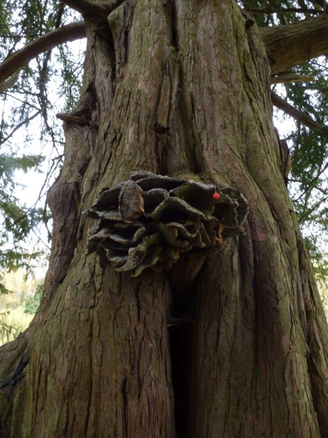
An old Laetiporus sp. on yew (Taxus baccata).

By virtue of the heartwood or ripewood being the least inhospitable (heartwood, in particular, is still often disgustingly harsh, as regards its environment), we can observe immediately some species-specific associations. For instance, the dyer’s mazegill (Phaeolus schweinitzii) will frequent gymnospermous hosts, such as the cedars (Cedrus spp.) and pines (Pinus spp.), whilst chicken of the woods (Laetiporus spp. – often L. sulphureus, though by all means not always, as we will see) will be found often on oak (Quercus spp.), sweet chestnut (Castanea sativa) and yew (Taxus baccata) – it can, indeed, be found on other hosts, as well. On the face value of things, these three tree species have seemingly little in common, though all three have extractive-rich heartwood that this genus can metabolise effectively. In terms of why the genus is referred to and not the exact species, Lynne is of the stance that what we term ‘chicken of the woods’ and default to as L. sulphureus is actually a variety of different species each with their own specialisations – perhaps even down to a specific host tree species (notably for yew, where the chicken species is most pertinently viewed as being a different one).

From a tree management perspective, Lynne then addressed the importance of the heart rotters – what is their impact? Put simply, they change the way in which we view the tree from a safety perspective; as in, when fruiting bodies of heart rotters are identified, if the tree is standing and there exists a target, management considerations are routinely entertained and sometimes the tree is felled. Additionally to this, however, we have other things to appreciate:

  • heart rotters do a great job at recycling nutrients, which can then be re-assimilated by the tree when they are uptaken back through its roots (including adventitious aerial roots) or the mycorrhizal fungi the roots associate with
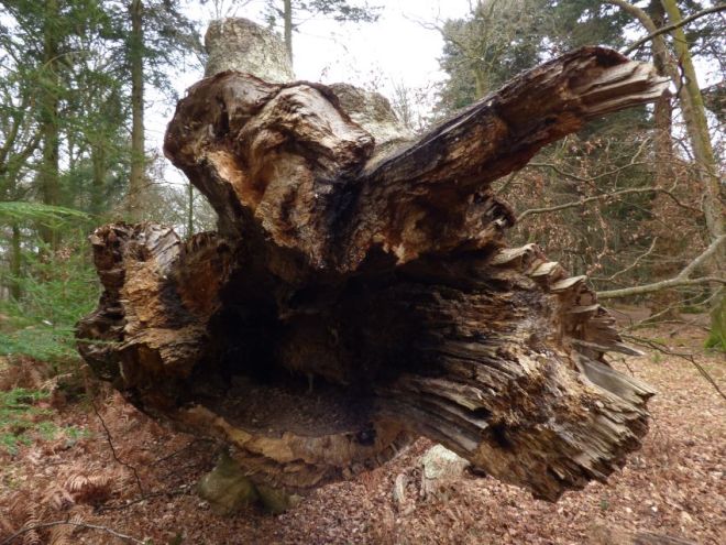
  • the wood qualities produced by heart rotters are ideal as habitat for saproxylic insects and nesting birds
phlebia-tremellosa-fagus-sylvatica-pollard-2
The heart of this beech has been hollowed-out by decay fungi. In the process, before its failure, what conditions did this decay provide for insects and, crucially, what habitat does it provide now?

Latent colonisers

Considered the specialised opportunists, such fungi are present within the sapwood or bark, as propagules (thick-walled resting spores known as chlamydospores). Biding their time until conditions are right, wherein the sapwood becomes dysfunctional through means such as wounding or drought (causing embolism), they are perhaps most acutely observed in the years after drought years where they can trigger the formation of strip cankers and resultant reaction growth by the tree (see the below photo). Thus, this year, in the UK, is one to watch out for, as regards such fungi (it might also explain why Kretzschmaria deusta was so abundant this year, given Ascomycetes love dry conditions, which prevailed last summer).

B nummularia Eutypa spinosa beech canker strip
A strip canker in beech caused by the latent fungus Biscogniauxia nummularia that has induced reaction growth, which can be seen cross-sectionally in the bottom right image).

At this point, a delegate enquired as to whether Massaria disease of plane (Splanchnonema platani), as an Ascomycete, could be prevailing in urban conditions recently, because of dry conditions (such as London, where it was been very severe these past few years but prior to that un-noticed). Discussions continued and Frank Rinn interjected to add his thoughts:

  • massaria progresses quickly in dry conditions
  • recent dry summers have allowed for massaria to thus progress very rapidly (killing branches in as little as three months)
  • the lowering of water tables in cities for the construction of basement levels of buildings has meant that plane trees can no longer tap into groundwater supplies
  • mature plane trees afford the best conditions for massaria; notably lower lateral branches, which are shaded from the rest of the crown and thus may be most prone to stress
  • there have been reports since 1903 from Croatia where large plane trees have shed branches and the massaria fungus was termed the “branch-cleaning fungus”
  • conditions are collectively ideal for massaria to become prevalent, as they stand

Reverting back to latent fungi, Lynne then mentioned that she considers fungi to be latent across a broad variety of trees. For example, the coal fungus (Daldinia concentrica), whilst found most often on ash (Fraxinus excelsior) can be isolated from the sapwood of a great range of different broadleaved tree species in the UK. It is, indeed, only when specific conditions arise that are preferable for this fungus that it begins to create mycelial networks – such conditions might not arise in particular trees, or may arise only after conditions suitable for other fungi have arisen and thus D. concentrica then has no capacity to colonise the substrate. Hence, ash remains the core host of this species, in the current climate. However, for the jelly ear fungus (Auricularia auricula-judae), which is also a latent fungus within the vascular system, having been found largely solely on elder (Sambucus nigra) in the 1950s, it is now found on over 20 host species – this marks a huge increase in host range, prompted perhaps by changing climatic conditions.

Wall V

The CODIT model, offered to us by Shigo, details four walls – as can be seen here. As mentioned by Frank Rinn, Shigo himself was considering the possibility of a fifth wall, though this never ‘made it into’ the model. However, Lynne argues that there is the potential for a fifth one, which hearkens back to what was discussed above, as regards rot within the heart of beech wood).

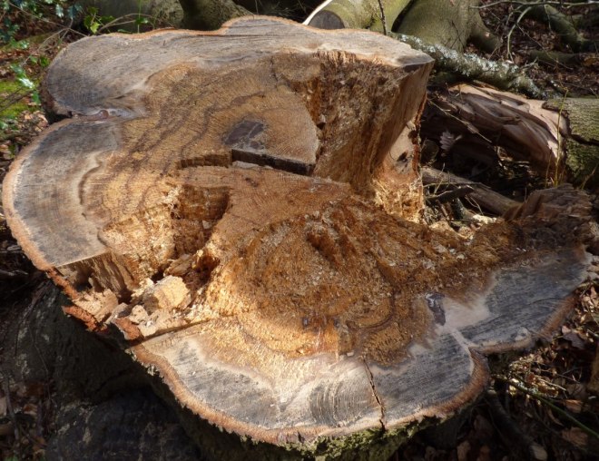
Specifically, whilst the barrier zone (fourth wall) is a zone laid down at the time of wounding by the vascular cambium, the dynamic responses by the tree that occur in real-time as fungal decay advances constitutes a distinction from this initial barrier. Indeed, as decay advances, living cells within the heartwood or ripewood (they do exist; though through mechanisms not fully appreciated, but thought to be associated with the rays running radially through the wood), in addition to the functional sapwood, will, in order to protect the sapwood and keeps its high-moisture quality intact, will plug woody cells beyond the current zone of decay with extractives and phenolic compounds – this will occur within the sapwood most often, though may also be able to occur in the heartwood around the regions where pockets of living cells exist. This response resultantly produces incredibly dense zones of wood that afford the tree’s sapwood a means of protection, which it would otherwise lack, assuming the barrier zone (Wall IV) failed to contain fungal decay. Of course, if this fifth wall fails, another will form, and so on and so forth.
Ganoderma pfeifferi beeswax beech failure 3
See the myriad of demarcations across this cross-section of a beech that failed from decay by Ganoderma pfeifferi, which suggest a dynamic fifth wall being effective.

Perhaps we will see this idea discussed more in Lynne’s re-write of Fungal Decomposition of Wood.

Fungal succession

When a tree decays, the fungi that initiated the decay process will not end it. This is because, much like all other ecosystems, as an environment changes those organisms that are best-placed to utilise it change as well. In this sense, fungi are no different – they succeed into a dynamic and ever-altering substrate (wood). Such a phenomenon can be so readily observed when walking into any woodland, when comparing the fungi on standing trees and those in early stages of decay and those much more heavily decomposed. In instances where an entire tree falls and stays largely intact, succession can be most acutely observed, as Lynne detailed with a little help from Ted Green and some research students.

The tree in question, a mature beech, failed and was left, in sections, for ease of its movement into an accessible place, to decay. Along the beech, it was found, through analysis of the wood in the laboratory and by presence of fruiting bodies, different fungi were observed colonising different parts at different stages of decay (as shown below). Such an observation does seem readily apparent, though to have it confirmed through scientific means affords us with an understanding that is more concrete than merely the anecdotal. Indeed, whilst Trametes gibbosa was not isolated from this beech, the presence of Bjerkandera adusta infers that, at some point in the future, T. gibbosa will be found – it parasitises upon the mycelium B. adusta, before then colonising the wood substrate itself. We are, in a sense, therefore, witnessing fungal warfare.
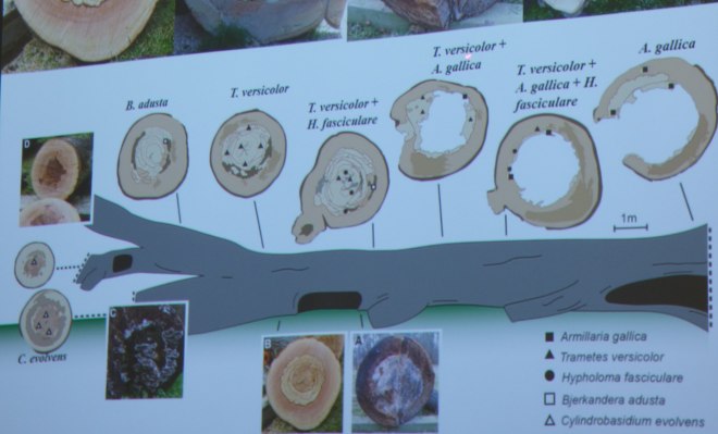
beech fungi succession analysis
The different fungi found at different parts of the beech at different stages of the decay process (open in a new tab to see this in a slightly larger size).

Delving further into the notion of fungal warfare, what is essentially meant is chemical warfare. Fungi synthesise and secrete enzymes, which they use principally to degrade wood, though that can also be used to defend territory or attack other fungi. The result of any fungal battle can be one of four things:

  • deadlock, whereby neither fungi gains any ground against the competing fungus
  • replacement, whereby one fungus loses its territory entirely by the other
  • partial replacement, whereby one fungus loses of some its territory to the other
  • mutual replacement, whereby the fungi essentially ‘trade’ places with one another and neither gains any net ground

So how does one determine the outcome of any such skirmish, you ask? Unfortunately, there are so many variables in play that even pitting two fungi against one another in a laboratory is only going to give a slight allusion to what really occurs, though there does nonetheless exist a limited hierarchy of combativeness from which we can assume who the victor will be, under most circumstances (see here at 25:09 timestamp).

Of course, even in assessing this we still have so many caveats to throw in. For example, where moisture conditions are drier because the wood is more exposed, Ascomycetes (i.e. Hypoxylon spp.) will have a better time in securing more wood substrate, as they operate effectively under dry conditions. Indeed, the wood qualities of the substrate itself will even play a role – did the tree uptake pollutants during its life or is it exposed to such pollutants currently, for example. More crucially, if a dead piece of wood (or entire tree) is standing and has thus been subject to relatively dry and exposed conditions suddenly falls to the woodland floor, those fungi reigning when the tree was standing will likely succumb to wood-decay fungi adapted to higher moisture levels and cooler more stable conditions.

The next part of this series will be a brief one on bacteria in wood, as discussed again by Lynne Boddy. I hope to have that written up in the coming few days.

Fungal succession and wood decay in living trees – a seminar report (Part II)

Burnham Beeches – old trees, wood decay and sun

A huge thanks to Burnham Beeches yesterday for hosting some of us for the day and showing us around the site. Below are some of the stand-outs from the day, which I am certain you will all appreciate!

The importance of functional units

As we can see in the below few images of a particularly striking beech pollard, very little of the structure of the tree needs to remain for the tree to persist as a living and functional organism. In this example, only one unit of vascularity supports a very small crown, though the beech is generally without significant fault. It could, potentially, persist in this state for many decades! Certainly, the two natural ‘props’ that support the crowd through a sort of tripod could, in their eventual failure, be the demise of this tree; assuming the functional unit cannot itself adequately support the crown. Depending on the rate of decay of this two ‘props’, this last vascular strip might (if decay is slow) – or might not (if the ‘props’ fail sporadically) – be able to lay down the necessary wood fibres for such mechanical support.

Fagus sylvatica functional unit pollard 1Fagus sylvatica functional unit pollard 2Fagus sylvatica functional unit pollard 3

Reduction work on lapsed pollards

There comes a point where one has to make a decision – for what reason is a lapsed pollard being managed? If it is to be managed for the provision of habitat then the major failure of the structure might not be an adverse occurrence (to a degree!), though if the intent is to retain the pollards for as long a period as is at all feasible then it might be necessary to undertake quite extensive reduction work, in order to reduce the mechanical loading upon the old pollard head. As can be seen from the below beech, heavy reduction work has taken place and the crown architecture / good number of ripe buds that remain below the pruning points will hopefully ensure that this lower crown will function very effectively. Of course, where lapsed pollards don’t have this lower growth then a heavy reduction might not even be possible, though where such low growth exists then it does provide for more effective means of management, with regards to reduction work of the crown.

Beech pollard crown reduction lapsed 1Beech pollard crown reduction lapsed 2

Submerged deadwood for reptiles

A terrapin uses a large section of a mostly-sunken stem for sunbathing, in the centre of a large pond. Indeed, this section of deadwood is an effective tool for the terrapin, which allows it to be exposed to direct sunlight and isolated from potentially aggressive mammals (that includes humans – seriously). Improving the texture and heterogeneity of this aquatic habitat with deadwood is evidently important, therefore!

Terrapin deadwood pond 1Terrapin deadwood pond 2

Artificial propping

As some of the old beech pollards are quite literally falling apart, safeguarding their structures against such cataclysmic failure is necessary, if their presence in the landscape is to be retained. For some, this involved reduction work, whilst for others it involves installing props to support either the enture tree or large / heavy parts of its structure. In the below two cases, we can see how props have been installed to stop the trees falling over completely.

Beech pollard propping artificial 1Beech pollard propping artificial 2

Wood-decay fungi

As you’d very much expect from a place such as this, wood-decay fungi are found in relative abundance. Beneath, the best examples are shown – this includes less common fungi, which we also came across during the trip; or less common associations, as you’ll see for one particular set of photos!

Fomitopsis pinicola (red-banded polypore)

Along the stem of a beech, this single bracket of a very infrequently found (in the UK, anyway) wood-decay fungus, the red-banded polypore, resides. Adjacent to a colony of Bjerkandera adusta and above extensive swathes of Kretzschmaria deusta, exactly to what degree this fungus has secured the wood substrate is unknown, though the good thing is that it has produced a fruiting body and in sporulating!

Fomitopsis pinicola UK Fagus sylvativa 1Fomitopsis pinicola UK Fagus sylvativa 2

Heterobasidion annosum (fomes root rot)

A common fungus but probably not one you see every day on hawthorn! Hidden beneath a branch ridden with Fuscoporia ferra (syn: Phellinus ferreus) and some leaves, a series of fruiting bodies were tucked away comfortably. Fungi love to throw curve-balls!

Heterobasidion annosum hawthorn Crataegus 1Heterobasidion annosum hawthorn Crataegus 2Heterobasidion annosum hawthorn Crataegus 3

Ganoderma pfeifferi (bees-wax polypore)

Sadly, the host beech had recently failed, due to the decay caused by this fungus. With respect to the rot induced, the failure was seemingly a brittle one and thus the failure can be attributed to a significant loss of cellulose. The cross-section of the failed region also yielded some glorious ‘rosing’ patterns, which is something that has been seen in other cases of failure as caused by this particular fungus.

Ganoderma pfeifferi beeswax beech failure 1Ganoderma pfeifferi beeswax beech failure 2Ganoderma pfeifferi beeswax beech failure 3

Fomes fomentarius (hoof fungus)

Found on both birch and oak, this species isn’t notably abundant in the south of England, where the pathogens Ganoderma australe / resinaceum / pfeifferi (in order of commonality) tend to be better suited. In the two instances shown below, fallen deadwood has provided the resource, which aligns with its colonisation strategy – that of awaiting stress / entire vascular dysfunction of an area or whole tree, before launching wide-scale colonisation activities.

Fomes fomentarius birch Betula 1Fomes fomentarius birch Betula 2Fomes fomentarius birch Betula 3Fomes fomentarius oak Quercus deadwood 1Fomes fomentarius oak Quercus deadwood 2

Daedalea quercina (Oak mazegill)

Found quite frequently on dysfunctional wood of oak, this instance has provided the best sight yet of this species. As you can see, an oak monolith is utterly littered with fruiting bodies, which is genuinely a spectacular sight!

Daedalea quercina oak monolith 1Daedalea quercina oak monolith 2Daedalea quercina oak monolith 3Daedalea quercina oak monolith 4Daedalea quercina oak monolith 5Daedalea quercina oak monolith 6Daedalea quercina oak monolith 7Daedalea quercina oak monolith 8

That’s all for today, folks!

Burnham Beeches – old trees, wood decay and sun

Fungi everywhere on a single declining beech pollard, New Forest (UK)

I was forunate to be able to spend some time in the New Forest yesterday, having driven back from Somerset after picking up a microscope (more on that, in due time). When last down there, which was during mid-summer, I spent a few hours sojourning around the Bolderwood / Knightwood Oak ornamental drive, with specific focus upon the myriad of mature and veteran beech pollards that dressed the roadside. One beech, even then, alluded to fungal parasitism, given its dire vigour and evident crown retrenchment (perhaps associated with ground compaction, given its close proximity to a car park and the Knightwood Oak). Therefore, I paid a visit to this beech, with the hope of finding some fungi – and I wasn’t disappointed!

I’ll actually be honest and say this beech is testament to the ability for the species to provide for many wood-decay fungal species. I really don’t think I have ever seen a tree more covered in fruiting bodies of many species than this one, and we’ll run through the suspected species below. First, we’ll look at the tree as a whole, however, and from the first image I don’t think there’s any debate over its poor condition. Granted, with the impending demise of a tree, weak fungal parasites and saprotrophs can enter, and this alludes to the cyclical aspect of energy transfer. In time, this beech will be the food for other plants and trees, though for now it’s fungal food.

fagus-sylvatica-various-fungi-wood-decay-whole
I wonder how many more years this beech has before its snatched from the throes of life! Probably not many.
fagus-sylvatica-various-fungi-wood-decay
The arrows relate to the various fungal species found. Working clockwise from the tip of the centre, I spotted what I suspect are Hohenbuehelia atrocoerulia, Chondrostereum purpureum, Mensularia nodulosa (confirmed), Exidia plana and Bjerkandera adusta.
fagus-sylvatica-mensularia-pleurotus-ostreatus-1
Here, behind a limb adorned brilliantly with one of the ex-Inonotus species, sit some fresh oysters (Hohenbuehelia atrocoerulea). Evidently, they are free from frost damage, suggesting they are probably only a few days old.
fagus-sylvatica-pleurotus-ostreatus
There’s also a younger set emerging just behind this cluster in the foreground!
fagus-sylvatica-mensularia-nodulosa-chondrostereum-1
Looking down the main stem, here we can observe how Chondrostereum purpureum and Mensularia nodulosa are inter-mingling. On the whole, it appears the Chondrostereum is more limited in its amassed substrate, if the presence of fruiting bodies are anything to go by – the ex-Inonotus species is abundant on the trunk and further up into some of the limbs.
fagus-sylvatica-mensularia-nodulosa-chondrostereum-2
In this image we can identify how the two species really do run right up to their respective thresholds.
fagus-sylvatica-chondrostereum-purpureum
For good measure, these are older sporophores of Chondrostereum purpureum. In their juvenile days, they’d have been far more attractive.
fagus-sylvatica-mensularia-nodulosa-1
Further round the trunk, we enter the sole territory of the Mensularia nodulosa.
fagus-sylvatica-mensularia-nodulosa-2
Angling upwards, the slotted nature of the tube layers becomes very evident.
fagus-sylvatica-exidia-plana-1
Down on one of the buttresses, this witches’ butter (Exidia plana) gets comfy amongst mosses. Note that it’s more likely to be this species of Exidia, as Exidia glandulosa is more often found on oak. To discern between the two however, you’d need to inspect some spores under the microscope.
fagus-sylvatica-exidia-plana-2
Looking more closely one can appreciate (I guess…?) why it’s called witches’ butter.
fagus-sylvatica-bjerkandera-adusta-1
And up on another limb, we have what is probably Bjerkandera adusta.
fagus-sylvatica-bjerkandera-adusta-2
It seems to be ejoying the decay column from the pruning wound and general dysfunction.
fagus-sylvatica-bjerkandera-adusta-3
There’s also some gilled sporophores in this one, which could potentially be Panellus stipticus, though they were too sparse and too small to see properly.
Fungi everywhere on a single declining beech pollard, New Forest (UK)

A wintry visit to Greenwich Park, London

Yesterday, as part of our monthly aim of visiting sites across the south east of England, a half-dozen strong group of arboriculturalists made the journey to London’s Greenwich Park – myself included. Indeed, as much of the park consists of deciduous specimens (principally, avenues of Castanea sativa and Aesculus hippocastanum), the park was rather bare in the foliage sense, though such barren canopies did allow us to appreciate the true magnitude of – most notably – some of the veteran sweet chestnuts. The frost-clad ground and crystalline sky provided a similar beauty, and thus we shall begin with one of the most iconic vistas from Greenwich Park – the city skyline.

greenwich-park-winter-city-view
As we stood adjacent to the observatory, we could admire – amongst the furor of tourists and scout groups – the sightly perverse beauty of a city. I say perverse, as such artificial and polluted landscapes don’t tend to suit those who don’t consider themselves urbanites, which includes myself.

Of course, we didn’t go there for the view, so let’s get into the main bulk of this account – trees and fungi. There’s no real order to how the below series of images rank, so don’t consider this post a chronological reflection of our trip!

Perhaps the best place in which to start the core section of this post are the huge sweet chestnuts, though we must begin on a rather sombre note. With a species of Phytophthora suspected on site and some of the older individuals exhibiting stunted and chlorotic leaf growth, there is a valid concern for the future of these veterans which is – without doubt – highly concerning. During the winter months, fully appreciating this contemporary issue is difficult, though we did spot some foliage on the floor that was certainly smaller in size than would be typically expected. Alas, this situation should not impact adversely on our admiration of these trees, and should in fact raise attention and draw intrigue to those within the industry and beyond, with an eye to ensuring we continue to care for the current and future populations of veterans. Therefore, promoting the Ancient Tree Forum and their most recent publication on ancient and veteran tree management is critical. And now, for some fine shots of various veterans!

castanea-sativa-veteran-uk-greenwich-1
This veteran sweet chestnut was the first one to greet us as we entered the park from the southern end. Not a bad induction!
castanea-sativa-veteran-uk-greenwich-2
As the city blocks paint the skyline to the right, we get a brilliant juxtaposition between the historic and the contemporary. In such a dynamic and ever-changing landscape such as London, this veteran sweet chestnut acts as a vestige of the old.
castanea-sativa-veteran-uk-greenwich-3
From another angle, the same sweet chestnut as above’s form can be more greatly appreciated. The helical patterns of the wood fibres and bark are as if they have been wound like rope.
castanea-sativa-veteran-uk-greenwich-4
This veteran has seen better days, though still stands proudly by the cafeteria. The ground beneath is woefully compacted, which must be having an impact upn the tree’s ability to function as a living being. Unlike the two shown above, it also doesn’t have a layer of mulch applied around its rooting environment.

Some of the veteran sweet chestnut we came across were also home to two annual common wood-decay fungi – Fistulina hepatica and Laetiporus sulphureus. Without doubt, the state of the fruiting bodies was not good, though when ravaged by time, wind, rain, frost and sun, to still even have a form is respectable! Certainly, a summer visit would have yielded a much greater haul of these two fungi on the sweet chestnuts, so a summer visit is probably on the cards.

castanea-sativa-veteran-fistulina-hepatica-1
One of Greenwich Park’s many veteran sweet chestnuts with an added extra – a small and rather weathered…
castanea-sativa-veteran-fistulina-hepatica-2
…you can see it…
castanea-sativa-veteran-fistulina-hepatica-3
…Fistulina hepatica! Picked off by parasitism before it reached a respectable stature, it still nonetheless produced a hymenium and thus likely produced spore.
castanea-sativa-veteran-fistulina-hepatica-4
A second sweet chestnut, this time slightly smaller, but again with Fistulina hepatica.
castanea-sativa-veteran-fistulina-hepatica-5
The state of it is, however, diabolical!
castanea-sativa-veteran-laetiporus-sulphureus-1
A smaller and thus younger sweet chestnut, in this instance.
castanea-sativa-veteran-laetiporus-sulphureus-2
It sports a fungal fruiting body, nonetheless!
castanea-sativa-veteran-laetiporus-sulphureus-3
A chicken of the woods, which is beaten and bruised.
castanea-sativa-veteran-laetiporus-sulphureus-4
Another smaller sweet chestnut, and another Laetiporus sulphureus.
castanea-sativa-veteran-laetiporus-sulphureus-5
Note how it emerges from behind a bark-covered area.
castanea-sativa-veteran-laetiporus-sulphureus-6
Again this sporophore is long beyond its best, though retains a little more dignity in the face of its impending crumble.

Away from the sweet chestnut, there was a variety of other large trees. Below, I share the ones that were home to fungi, through the identification of fruiting bodies. Absolutely, all trees on site are host to many species of fungi, though fruiting is not necessary in many instances, and it certainly costs the fungus energy to create and sustain. To begin, we’ll take a look at the ever-accomodating mature Robinia pseudoacacia in the park, which didn’t disappoint. In all, the population supported three species of wood-decay polypore, as we will see in the below images.

laetiporus-sulphureus-robinia-pseudoacacia-1
A very mature false acacia, with a very mature Laetiporus sulphureus fan on the main stem.
laetiporus-sulphureus-robinia-pseudoacacia-2
Well, sort of a fan – the remains of!
laetiporus-sulphureus-robinia-pseudoacacia-3
I imagine someone yanked this off, as it looks like a rather clean break.
laetiporus-sulphureus-robinia-pseudoacacia-4
Very close by, a second false acacia cradles another Laetiporus sulphureus.
laetiporus-sulphureus-robinia-pseudoacacia-5
Here, we can see how it’s at the base of the main stem, in place of higher up the structure.
laetiporus-sulphureus-robinia-pseudoacacia-6
This second one is far worse for wear!
perenniporia-fraxinea-robinia-pseudoacacia-1
A double-stemmed Robinia pseudoacacia, which was once at least triple-stemmed.
perenniporia-fraxinea-robinia-pseudoacacia-2
At the base, a senescent Perenniporia fraxinea and a cluster of broken active sporophores can be seen.
perenniporia-fraxinea-robinia-pseudoacacia-3
For good measure,here’s a better look at the entire bunch.
perenniporia-fraxinea-robinia-pseudoacacia-4
It’s a little disappointing that the fruiting bodies have been damaged, though that doesn’t stop them being Perenniporia fraxinea!
perenniporia-fraxinea-robinia-pseudoacacia-5
And a second example of Perenniporia fraxinea on this false acacia, too.
perenniporia-fraxinea-robinia-pseudoacacia-6
Right at the base, to the left.
perenniporia-fraxinea-robinia-pseudoacacia-7
This one appears slightly different to how it’d usually look (it’s not photogenic!).
perenniporia-fraxinea-robinia-pseudoacacia-8
Regardless, a showing of the trama reveals it as Perenniporia fraxinea.
ganoderma-australe-robinia-pseudoacacia-1
It looks like the park managers are aware of the decay on this Robinia, as it has already been pruned!
ganoderma-australe-robinia-pseudoacacia-2
If you look between the buttresses and into the basal cavity, you can spot a single Ganoderma australe. More were on the other side of the tree, though were old and worn.
ganoderma-australe-robinia-pseudoacacia-3
With the sun behind the camera, this southern bracket looks rather pretty.

Steering attention away from false acacias, I now turn towards a focus on the brown-rotting polypore Rigidoporus ulmarius. With both horse chestnut (Aesculus hippocastanum) and beech (Fagus sylvatica) on the site, the chances are that there would have been a few examples of this fungus. Indeed, there were, as we will observe.

rigidoporus-ulmarius-aesculus-hippocastanum-1
This first example, on horse chestnut, is an interesting one.
rigidoporus-ulmarius-aesculus-hippocastanum-2
It’s the return of the cavity-dwelling Rigidoporus!
rigidoporus-ulmarius-aesculus-hippocastanum-3
Away from the wrath of the elements, this sporophore doesn’t have the algal green stain atop and bathes in its own substrate.
rigidoporus-ulmarius-aesculus-hippocastanum-4
A cutting identifies this specimen as Rigidoporus ulmarius, with the cinnamon tube layer and brilliantly white flesh.
rigidoporus-ulmarius-aesculus-hippocastanum-5
The second horse chestnut sits in line for the toilets, patiently waiting for soneone to give it the 20p needed to get beyond the toll gate.
rigidoporus-ulmarius-aesculus-hippocastanum-6
If you want, you can even sit down to inspect this tree!
rigidoporus-ulmarius-aesculus-hippocastanum-7
This might well be this sporophore’s first season. I wonder how many more years it will see before it gets knocked-off or is aborted.
rigidoporus-ulmarius-fagus-sylvatica-1
Half way up this steep hill, a beech stands seemingly without significant issue.
rigidoporus-ulmarius-fagus-sylvatica-2
Oh, wait – here’s the issue!
rigidoporus-ulmarius-fagus-sylvatica-3
Is that a shade of green?
rigidoporus-ulmarius-fagus-sylvatica-4
From this shot, it looks most probably like Rigidoporus ulmarius. If so, we have two examples in one site of its cavity-dwelling abilities!

Greenwich Park also has a good number of large plane trees (Platanus x hispanica). The most abundant fungus on these trees was massaria (Splanchnonema platani), and there probably wasn’t a plane in the park that didn’t show at least some signs of its presence. However, it was the large plane with Inonotus hispidus that gained much of my eager attention, given I am not often around mature planes with extensive fungal decay.

inonotus-hispidus-platanus-hispanica-1
A rather lofty plane tree.
inonotus-hispidus-platanus-hispanica-2
As the crown breaks, we can spot a single Inonotus hispidus sporophore.
inonotus-hispidus-platanus-hispanica-3
Whether there is an old wound at or around this site is hard to say, though for this fungus to be able to colonise one would expect so.
inonotus-hispidus-platanus-hispanica-4
Perhaps an old branch stub above the fruiting body?

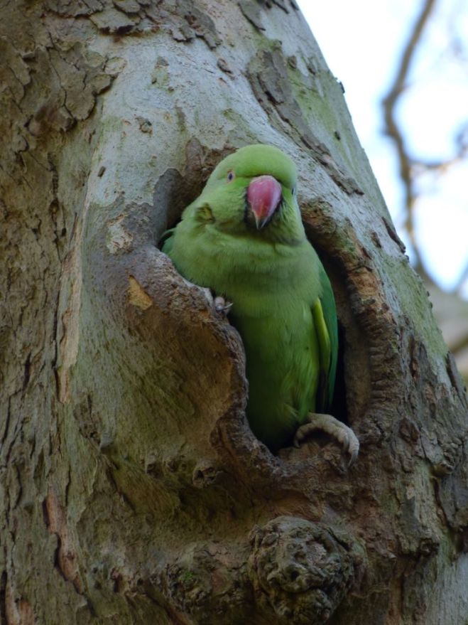
To round this post off, which has admittedly taken a long time to write, I’ll share some lovely images of a not-so-lovely bird – the parakeet (Psittacula krameri). Plaguing many of London’s parks and beyond, these things produce an utter cacophony and are certainly invasive, though one must admit that they are incredibly photogenic. Below, I share a few examples of where the parakeets were using cavities for shelter.

psittacula-krameri-london-tree-cavity-1
A horse chestnut monolith, seemingly vacant.
psittacula-krameri-london-tree-cavity-2
Wrong! Enter the parakeet(s).
psittacula-krameri-london-tree-cavity-3
This one stands proudly atop a pruning cut.
psittacula-krameri-london-tree-cavity-4
Along a plane tree branch, this parakeet appears to be guarding its abode.
psittacula-krameri-london-tree-cavity-5
“Oi m8, w0t u lookin’ @???”
A wintry visit to Greenwich Park, London

Meripilus giganteus finds from the field

For those of us in the arboricultural world, we are certainly aware of this wood-decay fungus. Its presence is often much maligned and associated with significant risk, and indeed in many an instance such concern is warranted. However, it can also act saprotrophically, decaying abscised or dead roots that provide no structural support. Therefore, caution is always to be exercised when making conclusions about trees with Meripilus giganteus, and Julian Forbes-Laird provides a good little write up on his views of the fungus here.

The purpose of this blog post is not, however, to pull apart the literature and assess what level of risk this fungus presents us with. Instead, I am here to show you a bunch of pretty awesome images, showing the host range I have personally seen Meripilus giganteus possess. Indeed, there are hosts not listed here that it is indeed found upon, though on a personal level seeing something first hand is much better than reading about it in a book and never actually getting to see what it is you have read. Get out and explore!

Acer pseudoplatanus (sycamore)

The only time I have seen this fungus on sycamore is on a stump – oh, how interesting…! To be perfectly honest, seeing it on a living host is far more exciting, though this does a good job of illustrating its ability as a saprotroph. Furthermore, it’s actually not at all bad as an example, and some oyster mushrooms even snuck in on the party – as did honey fungus on the root plate.

meripilus-giganteus-acer-pseudoplatanus-sycamore-1meripilus-giganteus-acer-pseudoplatanus-sycamore-2meripilus-giganteus-acer-pseudoplatanus-sycamore-3

Acer saccharinum (silver maple)

Again, this is an association I have observed only once. For the number of silver maple about and the fact that many of them are maturity, I’d expect for this association to increase in local abundance in the coming decade or two, though for now one single example suffices to prove the point – this association does happen! Unlike on the sycamore stump, it’s a single mass of fronds and has indeed peaked and is now therefore senescent. A shame I didn’t catch it a week or so before, but oh well – we need to be able to identify fungi in all their stages of life, when out assessing trees.

meripilus-giganteus-acer-saccharinum-silver-maple-1meripilus-giganteus-acer-saccharinum-silver-maple-2meripilus-giganteus-acer-saccharinum-silver-maple-3

Aesculus hippocastanum (horse chestnut)

Let’s be frank here – what doesn’t attack this poor species?! It is riddled with everything under the sun (I am surprised it hasn’t been attacked by ash dieback and Dutch elm disease…), and it being a suitable host for Meripilus giganteus is no different. The images below are from two separate trees, with one showing the near-primordial stage of fruit body formation, and the other demonstrating how bleedin’ sneaky this fungus can be. A master of subterfuge!

meripilus-giganteus-aesculus-hippocastanum-1meripilus-giganteus-aesculus-hippocastanum-2meripilus-giganteus-aesculus-hippocastanum-3meripilus-giganteus-aesculus-hippocastanum-4meripilus-giganteus-aesculus-hippocastanum-5meripilus-giganteus-aesculus-hippocastanum-6meripilus-giganteus-aesculus-hippocastanum-7

Carpinus betulus (hornbeam)

This is another association I have come across only once, and this hornbeam (which has seen far better days) happened to be laden with Ganoderma australe as well. Again, this one was caught a little late, though it once more outlines that Meripilus giganteus can surprise us with what it uses as a host.

meripilus-giganteus-carpinus-betulus-hornbeam-1meripilus-giganteus-carpinus-betulus-hornbeam-2

Fagus sylvatica (beech)

Yes, I have seen this association more than once – easily! It’s arguably fair to say that this is the most widely-known host for Meripilus giganteus, in the UK. Below are a selection of photos that show the better examples I have come across.

meripilus-giganteus-fagus-sylvatica-beech-1meripilus-giganteus-fagus-sylvatica-beech-2meripilus-giganteus-fagus-sylvatica-beech-3meripilus-giganteus-fagus-sylvatica-beech-4meripilus-giganteus-fagus-sylvatica-beech-5meripilus-giganteus-fagus-sylvatica-beech-6meripilus-giganteus-fagus-sylvatica-beech-7meripilus-giganteus-fagus-sylvatica-beech-8meripilus-giganteus-fagus-sylvatica-beech-9meripilus-giganteus-fagus-sylvatica-beech-10meripilus-giganteus-fagus-sylvatica-beech-11meripilus-giganteus-fagus-sylvatica-beech-12meripilus-giganteus-fagus-sylvatica-beech-13

Quercus robur (English oak)

This association is one I see a fair bit – not at all as abundant as it is found with beech, however. Typically, the oaks are at least mature and crown condition isn’t fantastic, though that’s nothing compared to the one I found on red oak below!

meripilus-giganteus-quercus-robur-english-oak-1meripilus-giganteus-quercus-robur-english-oak-2meripilus-giganteus-quercus-robur-english-oak-3meripilus-giganteus-quercus-robur-english-oak-4meripilus-giganteus-quercus-robur-english-oak-5meripilus-giganteus-quercus-robur-english-oak-6meripilus-giganteus-quercus-robur-english-oak-7

Quercus rubra (red oak)

For this one, I am just going to let the pictures do the talking. As far as I am aware, this oak is being removed, and rightly so!

meripilus-giganteus-quercus-rubra-red-oak-1meripilus-giganteus-quercus-rubra-red-oak-2meripilus-giganteus-quercus-rubra-red-oak-3meripilus-giganteus-quercus-rubra-red-oak-4meripilus-giganteus-quercus-rubra-red-oak-5meripilus-giganteus-quercus-rubra-red-oak-6

Sorbus aria (whitebeam)

Now this last one I wasn’t expecting! It’s very beaten and bruised and has succumbed to the elements a fair bit, but it is the only time I have ever seen this association and might well be the only time I ever do. The whitebeam is certainly mature, and the entire line are afflicted with one thing or another – this one has Meripilus giganteus, the next one has Ganoderma australe at the base, and another has its dead roots being colonised by Abortiporus biennis. The sorbi taking one for the fungal team here, evidently!

meripilus-giganteus-sorbus-aria-whitebeam-1meripilus-giganteus-sorbus-aria-whitebeam-2meripilus-giganteus-sorbus-aria-whitebeam-3meripilus-giganteus-sorbus-aria-whitebeam-4

Meripilus giganteus finds from the field

A visit to Wrest Park, UK

Last weekend I explored Wrest Park with a small group of other individuals, and we certainly saw plenty of interesting sights! For those curious, it is just north of Luton adjacent to a lovely old village called Silsoe, and for those arriving early you can even hear the bells of the nearby church ringing for Sunday morning service and explore the village to look at all the listed buildings.

However, as this blog is about trees, we shall keep it on all things tree-reated, and without further ado I’ll get into some of the features of this park that we passed by…

First of all, we came across this absolutely wonderful Wisteria sp. on the wall of the gardens. Without a shadow of a doubt, it is one of the largest examples I have seen, outside of the one at Kew Gardens. When trained well or given space to thrive, wisterias really do add a degree of formality and regality to a place, and one could hardly argue that the wall would have anywhere near as much character if this wisteria was absent!

wisteria-wrest-park-1
Here we can see how the wisteria envelops at least a segment of the much larger wall.
wisteria-wrest-park-2
And a great look at the stem morphology. The fluted appearance also gives this wisteria an old feel, which is probably because of its likely good age!

Just around the corner from this fine wisteria was a very sizeable cedar (Cedrus sp.). I recall it as probably being a cedar of Lebanon (Cedrus libani), though we’ll run with a species of cedar when noting my inability to remember specifics! I might add that this was a sizeable cedar, actually – it is now less so, but is certainly still a very impressive specimen. What is interesting about this cedar was the cavity three quarters of the way up what was left of the main stem, which sported some now rather senescent sporophores of the dyer’s mazegill (Phaeolus schweinitzii). The common name for this fungus comes from the fact that the fruiting bodies were used to make purple dye (a phenomenon not restricted to this fungus, as a very many fungi are used for dyes). Always be sure to check stem cavities for this fungus on conifers, and notably old cedars and pines, as they can yield some great fungal treasures!

phaeolus-schweinitzii-1
Wider than it is tall. I know they say that cedars flatten-out atop in maturity, though this one takes it a little too far!
phaeolus-schweinitzii-2
Of course, this cedar was a lot larger, though unfortunately lost its top – probably during high winds.
phaeolus-schweinitzii-3
But that isn’t a problem – the cedar still lives. And other things live within it, too – such as this dyer’s mazegill.
phaeolus-schweinitzii-4
A closer inspection with the camera reveals a small tier of fruiting bodies, and given the deep fissure they are emanating from it is certainly an historic wound and not a recent one.

Further around the gardens, we came across a huge copper beech (Fagus sylvatia ‘Atropurpurea’). As well as a discernible graft line that has begun to bulge, we can spot some highly distinct stretch marks on the bark. Evidently, the more recent annual increments have been quite marked in cross-sectional area, and the bark has therefore split to reveal a fresher bark layer beeneath. As we know, beech usually has a gloriously smooth bark, though in some instances that smooth bark is lost to a much rougher one. Is this a problem? Almost certainly not. It might however be associated with the graft point, if there are additional mechanical stressors acting on the area.

copper-beech-graft-stretch-marks-1
Here’s the copper beech, in all of its bare glory.
copper-beech-graft-stretch-marks-2
The trunk not only adopts a very stretched appearance but also has a pronounced bulge around the graft union.
copper-beech-graft-stretch-marks-3
A closer look just to appreciate the degree of bark stretching. Perhaps a visit to Boots would help this beech tree reduce the prevalence of the stretching!

As luck would have it, we then saw a lot of fungi. First came this horse chestnut (Aesculus hippocastanum) stump, which had on it not only Bjerkandera adusta, Chondrostereum purpureum and Flamullina velutipes, but many Mycena sp. on roots surrounding the stump. All, in this case, are saprotrophic fungi, meaning that they feast upon the abundance of dead wood, though in the case of Chondrostereum purpureum the mycelium will parasitise upon the mycelium of other fungi (a bit like how Trametes gibbosa will attack Bjerkandera adusta).

aesculus-various-fungi-1
A quite massive horse chestnut stump. In death, it still provides life for many species of fungi, as we can see…
aesculus-various-fungi-2
Some very fresh and guttating Chondrostereum purpureum. This species is very common on cut ends of fallen logs and stumps during autumn, and is able to parasitise upon other fungi. Thus, it is a fungus that is a secondary resource capturer, as it follows other fungi that came in before.
aesculus-various-fungi-3
More Chondrostereum purpureum alongside some rather old Flamullina velutipes.
aesculus-various-fungi-4
And a select few of the bonnets (Mycena sp.) surrounding the stump, which are very likely saprotrophs of dead roots.

Our walk then took us through the woodland garden, where Inonotus hispidus reigned supreme on some of the ash (Fraxinus excelsior). This woodland garden was rather peculiar, in the sense it was utterly laden with laurel (Prunus laurocerasus and Prunus lusitanica), huge yew (Taxus baccata), oaks (Quercus robur) with wonderfully straight boles, limes (Tilia x europaea) completely ravaged by mistletoe (Viscum album), and other tree species (such as ash, as was noted above). The growing amount of deadwood is certainly valuable for any invertebrates, fungi and bacteria, though the laurel does certainly require some management as it currently dominates the understorey.

inonotus-hispidus-fraxinus-excelsior-wrest-1
Obviously, as it is now late autumn, the leaves are off of the ash and the shaggy brackets are blackened (by-and-large). Here, we can spot one just before the stem break.
inonotus-hispidus-fraxinus-excelsior-wrest-2
Conveniently positioned beneath an old branch abscission point. Note the spindle-shape surrounding the old branch stub, which is the optimal way of ensuring there are no huge stress pockets in the area.
inonotus-hispidus-fraxinus-excelsior-wrest-3
This second ash is a little more sheltered but still doesn’t escape the ravenous hunger of Inonotus hispidus.
inonotus-hispidus-fraxinus-excelsior-wrest-4
Again, we can see it is associated with a wound. In this instance, the wound is a little larger in longitudinal extent. The area also sags slightly just beneath, indicating the white rot present (and thus delignification of wood) that is being caused by the mycelium of this fungus.

Also within this woodland, we came across a large oak that had lost its top during last winter. The debris now dressed the ground beneath, and it was not very surprising to see Bulgaria inquinans present on the fallen wood. This fungus is present as a latent organism within the vascular system of the oak, and upon death of a branch it quickly colonises (often quite brilliantly and over vast swathes of wood) to make use of the carbon readily available. In this example, the colonisation wasn’t so glorious, because the wood had been cut into smaller segments, though if left in tact the fungus would have produced black jelly discs across the entire upper stem.

bulgaria-inquinans-quercus-robur-wrest-1
A victim of the wind, though still alive. This loss of the upper stem might in fact allow the oak to live for a longer period of time, as it significantly reduces the risk of entire failure by the wind.
bulgaria-inquinans-quercus-robur-wrest-2
Some discs of Bulgaria inquinans, all of which are maturing or already mature.
bulgaria-inquinans-quercus-robur-wrest-3
Other equally mature examples on another piece of fallen wood. Absolutely abundant, as is to be expected.

After exiting this woodland garden, we were allowed access to a part of the site closed off to the public. Not only is this good because the area is often not trampled down, but what you can find in there away from the haunts of man is often a great treat, and we were not let down on this front, as you will see by the below images! However, before entering the gate, we stopped a beech (Fagus sylvatica) that had a great example of Perenniporia fraxinea at its base.

perenniporia-fraxinea-fagus-wrest-1
I suppose you could say it’s the guardian of the gate. It didn’t do a very good job though, as we had a key and the beech can’t actually move.
perenniporia-fraxinea-fagus-wrest-2
Here’s the sporophore, tucked away between two buttresses and covered by bramble a little.
perenniporia-fraxinea-fagus-wrest-3
Pay attention to the white spore beneath, which differentiates it from the slightly similar Ganoderma australe.
perenniporia-fraxinea-fagus-wrest-4
The brown context is another identifying feature of this fungus.

We then saw this fungus again, immediately upon entering the gated area. However, this time, it was on a host I had never seen it on before – hornbeam (Carpinus betulus)! And a great example it was, too.

perenniporia-fraxinea-carpinus-betulus-1
A rather contorted but very large hornbeam, with bark that one could almost describe as black locust-like.
perenniporia-fraxinea-carpinus-betulus-2
Around the butt were many sporophores of Perenniporia fraxinea, but this was by far the best one.
perenniporia-fraxinea-carpinus-betulus-3
Now here’s a postcard picture! Who wants to see a picture of a beach with palm trees when you can see this…!?
perenniporia-fraxinea-carpinus-betulus-4
And again a shot of the context.

Walking down alongside the lake we then came across a fallen weeping willow (probably Salix x sepulcralis ‘Chrysocoma’) that, upon its stem, was dressed with sporophores of the blushing bracket Daedaleopsis confragosa. This is rather common dead parts of willows (including Salix caprea), though generally the brackets are a little smaller and less sublime. However, in this instance, they provide us with a great example of how it looks.

salix-sepulcralis-daedaleopsis-confragosa-1
Willows doing what they do best – break!
salix-sepulcralis-daedaleopsis-confragosa-2
…and then providing great habitat for wood-decay fungi.
salix-sepulcralis-daedaleopsis-confragosa-3
This dead section of the stem is covered by over a dozen Daedaleopsis confragosa fruiting bodies.
salix-sepulcralis-daedaleopsis-confragosa-4
And when we look closer we can spot the brilliantly-textured upper surface. The pores below are between gills and pores, being quite elongated in nature.

So not to bore you all with fungi, I shall share one last example of Laetiporus sulphureus ssp. on yew (Taxus baccata). If you look around old yews you will often see chicken of the woods, though make sure never to eat it as it won’t do you any good and could end up with a visit to the hospital!

laetiporus-taxus-baccata-yew-1
If yew look closely…
laetiporus-taxus-baccata-yew-2
…you can almost see the tree weep at such a vile pun.
laetiporus-taxus-baccata-yew-3
This Laetiporus sulphureus ssp. even had a cherry on top. But don’t eat it…
laetiporus-taxus-baccata-yew-4
…because it’s blue and mouldy. And poisonous to some!

And now that I’m blue in the face from blaring out so many horrific puns, here’s a few images that will lighten up your day – lightning damage on oak!

lightning-damage-oak-1
Here we can see an old lightning wound that spans the entire length of the stem. Note the old ribbing to both sides and the central shallow fissure where the bolt probably tracked.
lightning-damage-oak-2
Another oak close by shows a similar style of wound, though less extensive in circumferential damage. Both oaks have survived, and long-term this damage might create some interesting habitat!

Well that is everything. I do suggest this site to those who are nearby, or want to make a trip at a weekend to explore a formal park. It is worth it, though please try to look beyond the huge yews that were topped to try and recreate a hedge! It isn’t easy, but I suppose the dead stumps make for some curious landscape features…

A visit to Wrest Park, UK

Dispersal and types of spores of Ganoderma lucidum (reishi)

So this is a very quick one but I wanted to share it as it’s actually really cool. As I am reading through Fungal Decomposition of Wood, on page 149 the authors detail that Ganoderma lucidum produces two types of spore from its basidia during its reproductive phase (where a fruiting body is present). Specifically, when the fruit body is young, thin-walled basidia are produced that germinate very readily on any suitable wood substrate that they may land upon (which is a slim chance anyway), though as the fruit body ages the type of spore differs to become much thicker-walled and – if it is to have any decent chance of germinating if it were to land on a suitable substrate – must first pass through the gut of fly larvae.

Of course, this differentiation in spore type (by where the former spore type is dubbed a ‘proterospore’) has implications for effective dispersal. The former thin-walled proterospores are to have a principal means of effective dispersion via air currents, and may indeed be released during the summer months when it is drier and therefore wind dispersal is more effective (assuming we apply a crude phenology to Ganoderma lucidum, and posit that a hymenium is produced and releases spores by summer time – late June through to late August). However, the thicker-walled spores produced in later stages of the fruiting body’s life (being an annual bracket, it will often senesce come late autumn, generally-speaking) would require either larval tunneling through the mature fruiting body and consuming spores along the way (which they then deposit elsewhere on possibly suitable wood substrates) or by larvae consuming spores where the spores were deposited by the air currents / other means. Larval tunneling could also take place after the bracket’s senescence, when some spores will still probably be ‘trapped’ within the tubes.

ganoderma-lucidum-carpinus-betulus-1
Three Ganoderma lucidum sporophores growing out from ever-so-slightly buried wood of Carpinus betulus. This old stump supported a good many more sporophores of this species.

If we are to accept that mature and active fruiting bodies are more likely to be host to tunneling insects, by virtue of: (1) the fruiting bodies being often sufficiently larger in size to allow for effective tunneling in a sheltered environment not at much risk of suddenly being aborted by the ‘parent’ mycelium and also possessing a greater supply of nutrients, and (2) because they have been in the environment for a longer period of time (thereby overlapping with ovipositioning of more insect species and generations), the second type of spore production actually makes logical sense. Furthermore, climatically-speaking, rain usually becomes ever more frequent as we approach late summer to early-mid autumn (when the Ganodermas are often still very much active, and the annual brackets of Ganoderma lucidum and Ganoderma resinaceum are typically fully mature – if not close to becoming so), and therefore wind dispersal perhaps becomes ever more challenging for polypores that depend upon dry conditions in which they can release spores effectively. In fact, and again it’s only my hypothesis, but as rain can drive spores onto and into the ground (as they are caught up in rain droplets, etc), the dispersal of these thick-walled spores might also provide them with the chance to be consumed by soil-dwelling insect larvae or ones that forage close to the ground, in place of upon fungal fruiting bodies or within their direct vicinity. When we note that Ganoderma lucidum can also fruit out from underneath the ground, it gives this theory time for a little more consideration. Thus, the potential scope of insect consumers increases quite extensively.

The caveat here is, as is most certainly evident, a lot of my own thoughts have expanded upon the initial statement in Fungal Decomposition of Wood. Thus, I may be spouting utter nonsense! Regardless, I hope it stimulates some thought, which is the most critical thing. As this book was written almost three decades ago and the research into Ganoderma lucidum spores was first published in 1982 by Nuss (cited below – it’s in German), there may have been developments since then that further explain this biological characteristic. At the very least however, this demonstrates that a spore is not just a spore, and that the mycological world is admirably complex.

ganoderma-lucidum-carpinus-betulus-2
Another series of Ganoderma lucidum upon the same Carpinus betulus stump, in the UK. This image was taken on 9th September 2016, as was the first image.

Sources:

Nuss, I. (1982) Die Bedeutung der Proterosporen: Schlußfolgerungen aus Untersuchungen an Ganoderma (Basidiomycetes). Plant Systematics and Evolution. 141 (1). p53-79.

Rayner, A. & Boddy, L. (1988) Fungal Decomposition of Wood: It’s Ecology and Biology. UK: John Wiley & Sons.

Dispersal and types of spores of Ganoderma lucidum (reishi)

Perenniporia fraxinea not on Fraxinus sp.

It’s fair to say that the epithet fraxinea is not indicative of the quite broad host range of this fungus. Granted, the same can be said about many other fungi, including Rigidoporus ulmarius and Pleurotus dryinus, and perhaps that’s one of the things we have to watch out for when it comes to identfying fungi on trees (and more broadly). Dealing specifically with Perenniporia fraxinea, which probably doesn’t have a common name, I want to share what other hosts I have come across that display signs of colonisation by this fungus (specifically, by identifying the fruiting bodies on the trunk and buttress area). This is, after all, an important aspect of tree management, and pin-pointing where fungi may occur outside of their suggestive range (by virtue of their epithet) is critical as a consequence. The mycological world loves to throw us curve-balls, and therefore we must be able to catch them when they are indeed thrown.

For the sake of ease, I have segmented the below pictures into headings detailing the different host species of Perenniporia fraxinea. I hope that some readers, either now or those who find this via a search engine in the future, find this of use – particularly those in the UK. However, before that, I shall list the host genera Ryvarden & Gilbertson list in their publication European Polypores (note this applies to all across Europe): Aesculus, CastaneaCeltis, Eucalyptus, Fagus, Fraxinus, Gymnocladus, Juglans, Olea, Malus, Platanus, Populus, Prunus, Robinia, Quercus, Salix, and Ulmus.

Acer pseudoplatanus (sycamore)

perenniporia-fraxinea-acer-pseudoplatanus-1
Only once have I come across Perenniporia fraxinea on sycamore and that was down in Dorchester, UK.
perenniporia-fraxinea-acer-pseudoplatanus-2
This sporophore as a bit of a mess, with regards to its morphology. Nonetheless, it had a hymenium, and was thus not an anamorph.
perenniporia-fraxinea-acer-pseudoplatanus-3
Thankfully, the other side of the same sycamore sported a more typical bracket-shaped specimen. Note the white spore around the bracket.
perenniporia-fraxinea-acer-pseudoplatanus-4
And a cross-section showing the distinctive context of Perenniporia fraxinea, including the fresh yellowish growth.

Aesculus hippocastanum (horse chestnut)

perenniporia-fraxinea-aesculus-hippocastanum-1
Again, only once have I observed this association. In this case, the horse chestnut was a monolithed specimen, and the sporophores were on the inside of the tree.
perenniporia-fraxinea-aesculus-hippocastanum-2
Trying to hide in frass and white-rotted woody material!
perenniporia-fraxinea-aesculus-hippocastanum-3
A cross-section, once again, reveals this fungus as Perenniporia fraxinea.

Fagus sylvatica (beech)

perenniporia-fraxinea-fagus-sylvatica-1
Three times have I witnessed this association on beech, and this was the most recent and the finest example of the bunch. Some great buttressing, too – as is so frequent on mature beech, which look like they’re essentially being ripped out of the ground in which they sit.
perenniporia-fraxinea-fagus-sylvatica-2
A closer look at this tiered specimen, complete with white spore dressing the buttress root. Upon cutting this one for a cross-section, it exuded a nasty ‘juice’.
perenniporia-fraxinea-fagus-sylvatica-3
Note the context, which is very distinctive of a specimen that has had its most recently growth ‘spurt’ mature – i.e. it lacks the yellow-orange rim.

Populus spp. (poplar)

perenniporia-fraxinea-populus-nigra-1
Both Populus x canadensis and Populus nigra ‘Italica’ are hosts I have seen Perenniporia fraxinea on within the genus Populus. Here, I share the better example of the two from the Lombardy poplar. A cracking buttress root there!
perenniporia-fraxinea-populus-nigra-2
On one side of the base we see this collection, all of which are laying down some very fresh growth.
perenniporia-fraxinea-populus-nigra-3
And on the other side some equally fresh growth spurts are taking place. Very photogenic!

Quercus robur (English oak)

perenniporia-fraxinea-quercus-robur-1
Having shared some other examples of Perenniporia fraxinea on oak, I wanted to share a newer example from last week. Three times I have seen this fungus on oak.
perenniporia-fraxinea-quercus-robur-2
Here we can see it’s certainly a sporophore with many years’ growth. This time around, a small layer underneath has been added.
perenniporia-fraxinea-quercus-robur-3
The white spore is so distinctive here, and the upper surface is covered in algae, etc.
perenniporia-fraxinea-quercus-robur-4
And a shot of the hymenium for good measure.

Robinia pseudoacacia (false acacia / black locust)

perenniporia-fraxinea-robinia-pseudoacacia-1
Robinia pseudoacacia is the second most common host after ash, in my experience (seen it a total of five times on this species). This is an example from earlier today.
perenniporia-fraxinea-robinia-pseudoacacia-2
Here we have a sporophore right at the base.
perenniporia-fraxinea-robinia-pseudoacacia-3
And some further up – albeit, much smaller, in this instance.
perenniporia-fraxinea-robinia-pseudoacacia-4
A closer inspection of two very good-looking sporophores.
perenniporia-fraxinea-robinia-pseudoacacia-5
And a closer shot of the one at the base – including the white spore once more.
perenniporia-fraxinea-robinia-pseudoacacia-6
And finally the context, as is typical of this fungus.
Perenniporia fraxinea not on Fraxinus sp.