When speaking of sapwood heterogeneity, it is meant that there is phenotypic variation within an individual (Karban, 2015). As trees will respond at local levels to different conditions of the external and heterogeneous environment (Schlichting, 1986), pathogenic invasion may not necessarily be ‘straightforward’. Areas exposed to different levels of particular ‘resources’ (water, light, nutrients) may differ in their cellular structure and contents, and thereby create a texture that differs throughout the tree and in turn increases sapwood complexity (de Kroon et al., 2005). This means pathogens may have a harder time invading into different ‘segments’ of the tree that have responded differently to other parts of the tree, due to localised environmental differences that occur on such a micro level (Adler & Karban, 1994).
How will the sapwood properties of this oak vary throughout its structure, and will such variation play a passive role in the tree’s defence against pathogenic invaders?
Earlier this summer (during late August), I noticed a developing Rigidoporus ulmarius sporophore between two buttresses of a hybrid black poplar. Because of the location of the tree, both in terms of the target area and its exposed setting, it was evident that some form of management would likely be needed. As a result, crown reduction works were issued during early autumn, and following a further inspection by myself in late October, I took a few more images for comparison’s sake.
My initial thought, having inspected the tree from afar, was that it was in good shape – the crown is clearly very dense, there is no indication of bark damage, limb failure, and so on.Peering through the longer grass at the base of the tree revealed something that changed my mind, somewhat – a developing Rigidoporus ulmarius sporophore. At this point, management considerations were particularly necessary. In this instance, a reduction was proposed.Returning two months later in late October, we can see how the form and colour of the bracket has changed. It is now slightly larger, and has the typical algal green atop.And here is the poplar following reduction. For me, the important thing is that risk is now temporarily reduced as a result of a reduction in dragging forces under wind loading, though future management needs must now be considered and undertaken on a cyclical basis.
Perhaps most renowned for the fact it was the cause of three deaths in Birmingham in December 1999, when the mature ash (weighing in at 15,000kg and estaimated to be 180 years old) that was host to this fungus failed and fell onto two cars waiting at traffic lights, Perenniporia fraxinea causes an intense white rot at the butt (lower stem and principal roots) of its host. However – on rare occassions – this fungus may be found further up the main stem, at sites of wounding.
According to current literature, this fungus can colonise – via either exposed sapwood or heartwood – species from the genus Fagus, Fraxinus,Laburnum, Platanus, Populus, Robinia, and Ulmus. It may more routinely, within the UK, be found colonising Fraxinus excelsior – particularly more mature specimens. Its preference for sporophore formation is however not fully understood, though I have seen it persist in an active state through to December.
As a result of its presence within the host, cavities may form (particularly in areas where decay is significant). Ultimately, the decayed wood from the lower stem and main roots is left increasingly prone to failure. In light of this, and in light of past events, it is advised that any tree colonised by this fungus is managed in a manner that reduces its risk to people and property. Of course, this may not mean pruning – the target zone, for example, could be fenced-off. This may be a particular avenue to pursue where the host tree is highly valued, or even as a means of studying the fungus.
Below are some images I have taken – all from mature ash trees close by to me – during the last month. Instead of describing its appearance to you, I would rather you look at some images.
At the base of a mature hedgerow ash, we can see a two-tiered arrangement. Note the white spore colour around the base of the brackets.A closer inspection reveals, more discernibly, the white spore colour on the ivy leaves beneath the brackets. Also note the resemblance to Ganoderma spp. (which have brown spores) and Rigidoporus ulmarius (which causes a brown rot).Factoring into the equation the fact my clipboard is A3 in size, we can really see how massive this bracket it. This ash was monolithed as a result of this decay, I strongly suspect. We can also see Daldinia concentrica above, which is a saprophytic fungus.Further around the base of the monolithed ash tree, we can see a sporulating bracket. The white spore colour is very significant here.Looking at a little closer at the underside of the bracket, the resemblance to Ganoderma spp. is understandable.A new bracket forming on another monolithed ash tree close by. Again, we can see white spores upon the ivy leaves, which distinguishes this from Ganoderma spp.
Disgustingly cliché title aside, I spotted something quite cool (well, there were more awesome things that I’ll be sharing from the visit) earlier on today whilst out inspecting trees in a cemetery – a Rigidoporus ulmarius bracket, detached from its host horse chestnut, masking a fresh growth right behind where it once sat.
So what is Rigidoporus ulmarius? It’s a fungus that, historically, would have colonised the butt of elms (given its scientific name), though as elms are less abundant in the UK thanks to Dutch elm disease, they can now more commonly be found colonising beech, horse chestnut, maple, oak, and poplar (amongst other species, I would expect). It’s rot type is a cubical brown one, meaning it dries out the wood by degrading its cellulose, and its presence can be of particular concern if extensive decay and / or cavities develop – this may lead to brittle fracturing at the tree’s base.
The horse chestnut this fungus has colonised can be seen situated next to a very infrequently-used car park and next to a well-used road.Sitting within some possibly very extensive reaction growth that, when hit with a sounding hammer, sounded ‘sound’, is the now-detached sporophore.Here we can really see how algae has colonised the sporophore’s surface – commonplace is such a sight with Rigidoporus ulmarius.And sitting directly behind this was a fresh growth that must have been 4cm in width at most. I put the detached sporophore back where I found it, masking this newly-growing one. Perhaps someone else will have a similar surprise in the future!
On Friday I made a post on Piptoporus betulinus and talked briefly about its colonisation strategy. You may recall that one of the images, which showed the underside of the bracket, had quite an interesting texture. As luck would have it, I think that I have just learned the reason (or one of the reasons) for this.
Here we see the bracket’s underside – the black dots may mark the entry points for boring insects, or perhaps even be small insects feasting upon the fungus’ flesh.
In his book Mycelium Running, Paul Stamets states that the ‘scent’ of the birch polypore attracts beetles that will, upon arrival, burrow into the spore-rich underside of the bracket and feast upon the inner flesh. As the beetles burrow in and feast, they are covered in spores. Upon leaving the birch polypore, the spore-laden insects will travel to another birch where they will bore into the wood and lay their eggs – all whilst depositing birch polypore spores.
The spores will then germinate and begin to form a mycelial web, which acts as a food source for the growing larvae. Because the mycelium induces a brown rot, the wood properties also change. In fact, as the wood properties change, woodpeckers are drawn to the tree in the search of grub to feed on. As the woodpekcers search for the beetle larvae, all whilst ‘damaging’ the wood during such a pursuit, they too act as a vector for spores – as does the woodpecker create conditions for other insects and birds to begin using this birch tree as a food source. Amongst all of the comings and goings of insects and birds, fungal spores hitch a ride, and the birch becomes a “launching platform” from which the birch polypore can continue its existence.
A couple of weeks ago I took a visit to Cambridge, not for the purpose of looking at trees, though nonetheless – as there were trees within the city centre – I couldn’t resist having a glance. Just as well I did, because I spotted a lovely Inonotus hispidus bracket on a weeping ash (Fraxinus excelsior ‘Pendula’).
I’m not quite sure what’s going on here, but there’s some absurd growth form on the main stem(s) – see the bottom part of the photo, and the area around the bracket. Perhaps an old branch tear out where the bracket is? No doubt that sapwood exposure has caused the fungus to colonise.The red circle shows the position of the bracket in this image. Unfortunately, I could not access the area to get closer shots.
Whilst Fistulina hepatica can be found upon its host wherever there is heartwood to be degraded, it’s normally found on the main trunk (particularly closer to the butt , because of the greater abundance of heartwood available). However, during the summer I spotted a small fruiting body up in the crown of a mature oak tree, on a branch with a diameter of around 15-20cm. So keep your eyes out for fungi where you may not expect them!
Here we can see the fruiting body emanating from a branch attached to a major limb.The red circle shows its location in this image, and demonstrates how high up in the crown it is.Having taken the branch attached off (there were visible signs of decay, stemming from this hollow that was likely an old branch attachment), we can see that the fungus was fruiting out of said hollow. Pulling off the bark revealed not only the brown rot associated with Fistulina hepatica, but also a stringy white rot of another fungal species.
Today’s journeys were fruitful on the fungal front: Daldinia concentrica, Fistulina hepatica (in a heavily rotted state), Ganoderma applanatum*, Ganoderma australe*, Laetiporus sulphureus (again in a rotted state, though also blitzed by an animal), Piptoporus betulinus, Porodaedalea pini, and Pseudoinonotus dryadeus were all finds. (* I strongly suspect both Ganoderma spp. mentioned were identified, given very distinct morphological differences between the sporophores). The topic of this post will be Piptoporus betulinus exclusively, as the sporophores I saw really raised my interest (in part, down to my newly-acquired Swiss Army Knife and its dissective powers).
Birch polypore really isn’t an uncommon site where birch can be found in woodland – rarely do I visit a woodland with birch and not see either active or more desiccated examples. However, it’s the colonisation strategy of the fungus that really enthralls me. Spores will gain entry via stem injury, and then attack the tree’s sapwood and heartwood once once the host is under stress – in woodlands, this may very well be induced by a birch’s poor ability to compete for light with maturing (typically) later-successional and / or slower-growing species such as oak, beech, chestnut, hornbeam, and ash. Once the host birch is stressed, the fungus will attack, creating a (quite wonderful in appearance) brown rot, and the stressed host will likely die very swiftly (if not fail a few metres up from the base). Birch polypore will then persist as a saprophyte – this is when, for me, the fun happens. Just look at the below photos.
Oh look – it’s a dead birch. And what’s on it? Birch polypore!Looking upwards from the ground, we can identify five different sporophores (at all sides of the circumference) on this dead host from this angle.Nearby, another dead birch acts as a host. Notice the trunk has fractured half way.Taking a step back to view the host, we can see that the entire remaining stem is colonised – all the way down to the base. And we can now see that this dead birch failed initially up high on the stem, and has since (or even at the same time) failed beneath.Given their local abundance, I took one back for a closer inspection. Here we can see the texture atop, and the dainty stalk. …and the texture below. I can see a resemblance to desiccated Laetiporus sulphureus. I then cut open, for the first time, this fungus’ sporophore. If I’m honest, I didn’t expect what I saw – an immaculate and pure white cross-section (the marks were from my blade), with but a tiny underside where the spores are released from. Pretty awesome, if you ask me!
Stopping off for a quick break during work today, I parked under an oak that has always been of interest to me. Situated in the main car park for a large amenity park, not only is its mere existence intriguing (in many instances, such trees would likely have been removed), but its appearance sparks the imagination – amongst other qustions, what has this tree endured, and what ecological benefits does it provide?
Still clinging on to life, this oak certainly has a lot of amenity value. I am certain that many park users walk by this tree and take notice of it. The rich organic matter pouring out of the base can also be seen, courtesy of fungal (and insect) decomposition.
As a matter of fact, this little critter could be seen fruiting within the tree itself during the summer.
Can you see it? Look towards the centre of the image.Here we can see a tiny Fistulina hepatica has developed. I have seen such tiny sporophores of this species a few times – such an interesting fungus, morphologically-speaking.
Farmers may likely resort to placing large pieces of timber at the boundary of their land, in an attempt to stop vehicles driving onto their property and using it for either a temporary home (as travellers may do), or otherwise. Not only is such a tactic incredibly effective at halting vehicular trespass, but also more ecologically beneficial than constructing, for instance, a man-made fence.
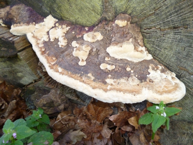
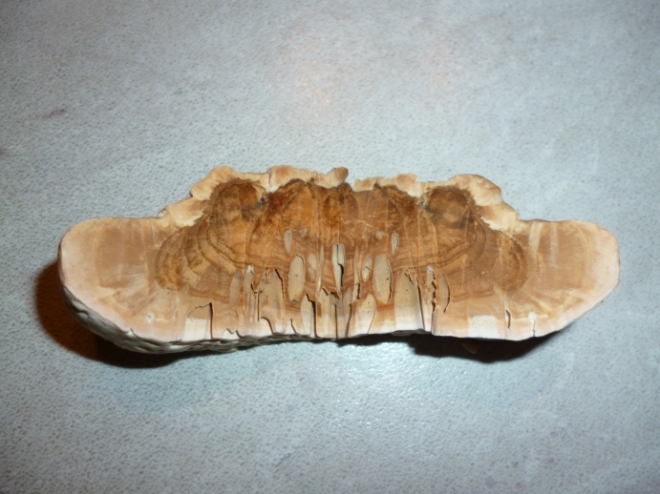
On this oak log, we can see that Daedalea quercina (oak mazegill) has colonised and subsequently produced a sporophore. I first noticed this earlier in the year during an ‘inactive’ phase, though recently it has sprung into life again. I therefore took the opportunity to take some photos and a small sample, of which the results are below.
The oak mazegill, according to Mattheck et al. (2015), can be both parasitic and saprophytic, though I have only ever seen it act saprophytically. It induces a brown rot of the heartwood by preferentially degrading the cellulose, and is principally found on oak (though sweet chestnut may also be a host).
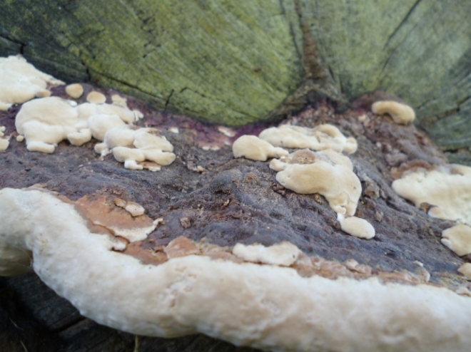
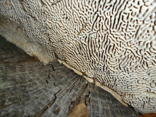
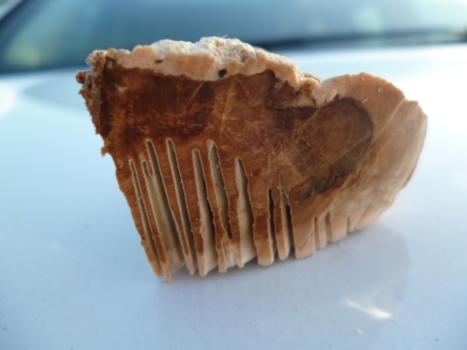
A view of the entire bracket, which measures around 30cm in width, upon the end of an oak log.A closer inspection reveals the wonderful texture of the bracket.Here we can see where this fungus gets its common name.A small sample taken from the bracket reveals the inner workings of the bracket, and also shows how much new growth has been laid down in this growing period (note the sudden transition between dark brown and beige).Cutting the sample in half and folding it out produces a very lovely symmetrical image, if nothing else!